How to See Thoughts: Non-Orthodox Applications of Magnetic Resonance Imaging
Today magnetic resonance imaging (MRI) is used not only for diagnostics but also for mapping the functional state of neuronal networks, allowing one to literally see the brain working in real time. MRI has enabled the development of a game biofeedback technology based on the natural self-regulation of human bodily functions. The unique computer games designed by Novosibirsk researchers teach the user to “direct” a virtual game plot via voluntary changes in his/her physiological characteristics, such as pulse, skin temperature, brain electric activity, etc. The games are applicable to a wide range of therapeutic and rehabilitation tasks, including the assessment of the current psychophysiological state of a person. In addition, these playing activities are themselves a good antistress factor. However, most importantly, this technology uncovers our potential resources, which we are yet unable to use in our daily life
Until recently, the basic data on the brain functions were obtained only from indirect sources, namely from experiments with animals; from observations of the patients with affected brain areas that appear as paralyses or speech and memory disorders; from neuropsychological testing; from open brain surgery that allows the neurosurgeon to observe responses to particular stimuli; and, finally, from the recording of the brain electric activity. However, the data generated by such approaches do not permit any description of how the brain works when solving a particular problem. Only the advent of functional magnetic resonance imaging made it possible to directly observe the dynamics of the brain cognitive activity
The hypothesis postulating a correlation between the brain blood supply and its activity became widespread as early as the late 19th century thanks to C. Sherrington, an outstanding British physiologist. Many years later, this correlation was proved by radiography methods, which confirmed a direct dependence between metabolic processes in certain active brain areas and the rate of oxygen delivery to these areas.
Over two decades ago, a team from the AT&T Bell Laboratories described the principle allowing for a real-time activity visualization in brain areas, using magnetic resonance imaging (MRI), in which the image contrast is determined by the degree of blood oxygen saturation (Ogawa et al., 1990). This principle formed the foundation for the so-called functional magnetic resonance imaging (fMRI), which is a dynamic examination of active brain areas at the moment they fulfill their function; for the first time this technology was tested in humans two years after the first publication.
Oxygen as a marker
Activation of the brain area is always associated with oxygen consumption; correspondingly, it entails acceleration of glucose metabolism and transformation of hemoglobin molecules, oxygen suppliers in our body, when oxyhemoglobin with reversely bound oxygen is converted into deoxyhemoglobin (reduced hemoglobin).
The key factor for MRI is the distinctions between the magnetic properties of different hemoglobin species. In particular, oxyhemoglobin is a diamagnetic, i.e., the substance creating a magnetic field in the direction opposite to the external magnetic field. On the contrary, deoxyhemoglobin (reduced hemoglobin) is a paramagnetic, which creates the magnetic field of the same direction as the external field. The value of the MRI signal depends on the amount of deoxyhemoglobin in tissue: the higher its concentration, the lower is the signal. The characteristic determined by the ratio of these two hemoglobin species and depending on the oxygen level in the blood is referred to as BOLD (blood oxygenation level dependent).

Then the generated signal is converted into the so-called T1-weighted image (T1 is the time required for two-thirds of the protons to return to their initial state). The output image will be different for individual tissue types, for example, for healthy and damaged ones.
State-of-the-art MRI technologies make it possible not only to get high-quality images of various internal organs, but also to study their functions. Thanks to the absence of ionizing radiation, this method has no limitations for application and can be used repeatedly
The more actively working is the brain area, the more oxygen it consumes. When an acting neuronal ensemble is being formed, an increase in the local energy consumption leads to an increase in the deoxyhemoglobin (a paramagnetic) concentration in the very first seconds; the next response comes from the vascular system as an increase in the blood supply and filling of the brain tissues caused by the growth in blood circulation volume and rate.
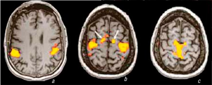
Correspondingly, the relative value of the MRI signal may serve as an activity measure for the brain areas. Moreover, the MRI results for the visual cortex of an ape’s open brain under the EEG control suggest that the MRI signal is a linear response to the electric activity generated by the acting neuronal ensemble (Logothetis et al., 2002).
Thus, the functional MRI focused on detecting the BOLD effect is currently the optimal tool for mapping neuronal activity or, more precisely, the functional state of neuronal networks, which are the base for visualizing our thoughts and ideas. In other words, fMRI literally allows us to see, in a real-time mode, how our brain solves problems.
Power of thought
fMRI is closely associated with a neurobiological technology referred to as brain—computer interface, a kind of computer symbiosis (Kaplan, 2005, 2012; Chernikova et al., 2010). Here we mean the possibility of getting an image of a stable “pattern” of the brain bioelectric activity by electroencephalography, affiliating this pattern to the functions of brain structures and formation of new neuronal ensembles there. Note that the electroencephalogram in this case is not only a source of information about the events in the brain; these data may be also used as a feedback signal for the voluntary self-regulation of the body functions.
As early as at the end of the 19th century, the French neurosurgeon Paul Broca described speech problems caused by the damage to a certain area in the left hemisphere. His work laid the foundation for numerous studies devoted to the clinical analysis of the brain’s language organization and its impairments. Thus, determining speech development, namely the localization of the speech center within the corresponding brain zones, has become one of the largest application areas for fMRI.The data on the location of speech (literal, semantic, and syntactic) zones in the brain is now used in neurosurgery. This involves a pre-intervention detection of the particular cortical areas in the patients with different cerebral affections, which are prohibited for the surgeon’s knife. Today, fMRI is almost the only technology that can find such a frontier zone
Although neurobiology is an independent science, it emerged as a “social product” for seroiusly disabled people; it gives a chance to wheel-chaired people deprived of motor skills to control artificial appendages such as “a mechanical arm” (Hochberg et al., 2012).
One of practical applications of neurobiology is neurobiofeedback—a non-drug technology based on the principles of the above mentioned feedback, the phenomenon underlying the self-regulation mechanism. This technology uses the idea that a person can learn to voluntarily control unconscious physiological characteristics such as the pulse rate and parameters of the brain electric activity rhythm.
As early as in 1958, Joe Kamiya was the first to describe the human ability to purposefully change EEG parameters; this ability was studied in order to control the functional state of a patient’s brain and to change trends of mental development. Further studies demonstrated the amazing capacities of our brain for internal rearrangements not provided by Mother Nature. Neurobiofeedback appeared to allow people developing self-regulation skills that did not exist earlier, to form new skills, and to “awake” dormant brain structures. In this situation, fMRI makes it possible to visualize real-time and spatial dynamics of the brain function.
From a practical standpoint, of special interest is the technology for the so-called game biofeedback, when a person learns to “direct” a virtual game plot via voluntary changes in his/her physiological characteristics such as the cardiogram, pulse, skin temperature, and brain electric activity.
Mastering oneself
In the context of neurobiology, a game is a psychological reality with a large number of nontrivial situations unresolvable by a stereotypic behavior pattern. A computer player gets used to traveling from one virtual world to another, rapidly adapting to new virtual realities based on his/her personal preferences.
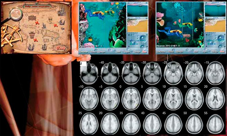
While playing a game, the brain actively functions in order to determine the way of actions that is the most beneficial for the moment. If the player uses biofeedback, after acquiring the self-regulation skills, he/she can control this process, since an adaptive feedback makes it possible not only to see and play over different behavioral strategies, but also to estimate their efficiency. In this sense, this technology is a powerful mechanism allowing people to learn new behavior stereotypes.
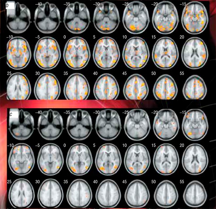
When practicing a game biofeedback, the player takes an active role in the therapeutic (correction) process and acquires new skills
The Institute of Molecular Biology and Biophysics, Siberian Branch of the Russian Academy of Medical Sciences, in collaboration with and using the facilities of the International Tomography Center (Novosibirsk, Russia), conducted an experiment on neurovisualization of “voluntary” control of a virtual game plot with a cohort of young men.
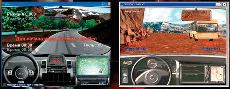
The participants played the game Vira!, which involved searching for underwater treasures. Each participant, placed in a ring-shaped scanning magnet, controlled one scuba diver going to the bottom. The player’s speed was directly determined by the heart rate: the lower the heart rate, the quicker was the diver. During the game, the heart rate data were shown as a graph on the screen. In order to win the game, the player had to learn how to control the heart rate mentally, that is he had to develop the skill of slowing down his heart rate.
The game results showed six different behavior patterns; the leading self-regulation strategy was determined for each one.
In particular, the participant using the trial and error strategy initially made several unsuccessful attempts but eventually achieved the goal. The participants using this strategy focused on controlling the game result rather than on their physiological parameters, i.e. the pulse. The pendulum strategy was characterized by an alternation of successful and unsuccessful attempts while the consistent education strategy, by the successive improvement of the result from one attempt to another.
Analysis of the experimental data demonstrated a certain sequence of the emerging and developing of the activity zones in the brain of the participants. The peak of the competitive game plot was observed at the fourth—sixth attempts, when an ever increasing number of newly formed neuronal ensembles were involved into the efforts to win.
Interestingly, the new zones of this activity were also detected in the cerebellar structures. Analysis of their formation dynamics suggests that the cerebellum of our brain not only regulates motor functions, but also modifies cognitive functions via regulating the brainwork rate, strength, rhythm, and accuracy. In this process, the program of cognitive operations is being successively implemented in the mode organized by the adaptive feedback.
This is how the “roadmap” for cognitive control of the Vira! game line was formed according to the trial-and-error strategy, the most widespread self-regulation pattern.
A lie differs from the truth
The virtual reality represented as a competitive game controlled by the voluntary regulation of a physiological characteristic gives a person a unique chance to exercise the specific behavior features that are usually blocked. In this sense, not only a virtual game, but also any game training allows us to detect the concealed abilities which we can successfully use in real life.
In this context, of special interest are the results of the game experiment conducted at the International Tomography Center, which involved the so-called “simulation,” i.e., false, biofeedback in addition to the “real” one; in other words, when the game plot developed randomly and was completely independent. The participants themselves did not know that the real feedback was absent in one of the experimental series.
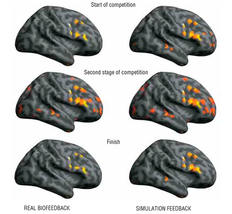
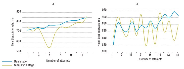
The participants of this experiment fell into two groups according to the effectiveness of their game results. The first group displayed more efficient self-regulation strategies in the presence of real feedback than in the presence of the false biofeedback. Moreover, even in the absence of real feedback, some participants, after several unsuccessful attempts, succeeded in slowing down their heart rate.
The second group displayed a less efficient self-¬regulation strategy: even at the stage of “real” feedback, these participants reached the goal only partly. When there was no feedback, their search for solution was intensive and “chaotic,” which was manifested as a widened range of their pulse. Nonetheless, both groups displayed a higher self-regulation efficiency in the event of real biofeedback as compared with the simulation, proving that our brain is able to differentiate between the “truth” and a “lie.”
Note that both real biofeedback and its simulation were accompanied by a distinct dynamic pattern of the activity in several brain structures, appearing as changes in the activation space and redistribution of activity zones. Actually, all the brain cortex was involved in the process; moreover, most of the cortical zones involved in the simulation and real trainings overlapped and displayed the maximum activation values in both cases. However, in the case of simulated feedback several brain structures were considerably stronger activated as compared with the real feedback situation: new neuronal ensembles were formed in the cerebellum, in the lateral occipitotemporal gyrus, and in some other brain structures.
WIN-WIN GAMES A team from the Institute of Molecular Biology and Biophysics (Siberian Branch, Russian Academy of Medical Sciences, Novosibirsk) and the Computer Biofeedback Systems research and production company (Novosibirsk) creates a unique product, computer games with a competitive plot that is controlled by physiological characteristics of the human body (temperature, pulse, breathing, and brain and muscle biocurrents).The technology of computer game biofeedback is based on the natural self-regulation mechanisms of the human body functions. The competitive character of such games removes the monotony of the learning procedure: an entertaining plot motivates the participant, causing emotional interest and thus making the learning of self-regulation skills more efficient. Since victory requires for the participant to make nontrivial decisions, such a game may be regarded as creative learning, the attraction of which consists in unpredictability of the final result. Since each game attempt is based on the results of the previous one, the game biofeedback ensures self-improvement of the participant as well as encourages a search for new efficient self-regulation strategies. Since the player is motivated by the desire to win, he/she has to keep within the frame specified by the game and keep calm.
The games based on the biofeedback technology can solve a wide range of therapeutic and rehabilitation problems. They help to assess the current psychophysiological state of a person; in addition, such playing activities are themselves a good antistress factor. But the most important thing is that this technology allows us to discover our potential resources, which we are yet unable to use in our everyday life
An attempt to describe the most general “route” in activating the brain structures during the game shows that wide cortical areas are the first structures activated after the game starts and that the “cognitive route” ends in the cerebellum. Successive involvement of the brain structures into organizing new neuronal networks during such a virtual training results in acquiring a new skill and its subsequent fixing in the brain. In this sense, the studies of this type fall into a new trend in the development of social environment referred to as “gamification.”
Efficient or fair?
Psychology is among the most promising fields for the application of neurovisualization provided by fMRI, because this research area almost does not operate with the localization (in an anatomical sense) of cognitive functions. Indeed, psychologists acquire most of the information about “territorial reference” by speaking with the patients with instrumentally diagnosed local brain lesions or with implanted intracerebral electrodes.

A team of American researchers once attempted to answer the question about the localization of the brain structures capable of classifying such cognitive categories as equality and efficiency (Hsu Ming et al., 2008), or, in other words, the structures aimed at solving the long-lasting dilemma of whether one should act efficiently or fairly.
In one of the game experiments, participants were “driving” a truck that carried foodstuff for starving people in a South African area. It was set that if a player kept strictly to the rules and gave an equal share to every starving person, part of the food would spoil on the way. However, if half the starving people were deprived of their shares, the food loss would decrease considerably, but fewer people will be fed. So, what is to be done? Should part of the food be sacrificed or should half the needy be left without any hope for help if the players made a “reasoned” choice?
Three brain structures appear to be involved in the emotional estimation of the “efficiency,” “justice,” and “overall benefit” of the decision made. The brain part referred to as the putamen is responsible for efficiency; the insular cortex acts for justice; as for efficiency and inequality together, i. e. usefulness, it is evaluated by the septum.
These results agree with the data available that it is the above-mentioned brain structures that act as integrators of various psychic “variables” in delivering the final “socially-oriented” judgments and estimates. Presumably, the final solution to an ethical problem is found by comparing the signals from different sources and by collating them with retrospective experience; other brain areas are also involved in the cognitive process.

The number of published papers on different basic and applied aspects of functional magnetic resonance imaging and brain computer interface is steadily growing (mainly abroad); there are practically no Russian papers in this list. The development of corresponding technologies simultaneously opens several promising applied areas. For example, now it is posible to monitor specific blood circulation features in an activated brain segment, and in particular, to monitor certain brain structures in the case of impaired blood circulation in the brain (a stroke) or when selecting optimal vascular drugs.
Great opportunities are offered by the development of cognitology, an area in neuroscience that deals with the basic mechanisms of the brain operation, namely with “mental strategies” and their localization, dynamics, as well as application and improvement in everyday life. The so-called “interactive stimulation” makes it possible to form a training (remedial) feedback directly via the “engaged” brain structures. For example, visualizing the cingulate convolution or hippocampus gives a chance of a “direct conversation” with the brain.
Functional magnetic resonance imaging is a powerful tool giving a qualitatively new understanding of the brain organization and of specific features of the human and animal higher nervous activity. Introduction of fMRI technologies into various spheres of human activity – neuromarketing, professional casting, efficiency assessment of educational programs, lie “detection”, etc. – will have a great impact on the further development not only of neurosciences, but also of society as a whole.
References
Kaplan A. Ja. Nejrokomp’juternyj simbioz: dvizhenie siloj mysli // NAUKA iz pervyh ruk. 2012. № 6 (48). (in Russian)
Shtark M. B., Korostyshevskaja A. M., Rezakova M. V., Savelov A. A. Funkcional’naja magnitno-rezonansnaja tomografija i nejronauki // Uspehi fiziologicheskih nauk. 2012. T. 43, №1. S. 3—29. (in Russian)
The photos are by the courtesy of M.A. Pokrovskii


