L’Oreal-UNESCO Scholarship: Beauty and Intellect Make a Perfect Match
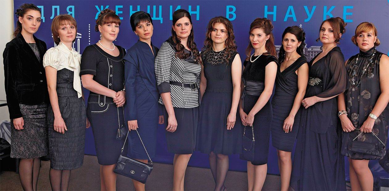
Yet again, the national L’Oreal-UNESCO scholarship “For Women in Science” worth 350,000 rubles was awarded in Moscow on the Day of Science (November 10, 2010). The national L’Oreal-UNESCO scholarship scheme now functions in 45 countries. In Russia, the competition, co-sponsored by the Russian Academy of Sciences, has been held since 2007. Applicants can be young women (Candidates of Science under 35 years old) engaged in pursuing research in physics, chemistry, medicine or biology at Russian research institutes and universities. In contrast to other competitions, participating in this one is easy: you should just fill out a short form. In 2010, four hundred applications from different cities of Russia were received. Ten participants were chosen to be the prize winners. All of them are successful scientists, well-known specialists in their fields, leaders of research groups and laboratories.
This time, the Siberian Branch of the Russian Academy of Sciences (SB RAS) has given Moscow a good run for its money: four award winners out of ten are from Novosibirsk Research Center SB RAS. Today, we are presenting two Siberians: a specialist in X-ray structural analysis Eugenia Peresypkina, given the highest rating by the award committee, and Svetlana Tamkovich, engaged in the development of promising methods for cancer diagnostics
Crystallochemical mosaic
The main task of empirical crystal chemistry is searching for general regularities in the crystal structure. These studies are extremely important because most solids surrounding us are crystalline.
In contrast to other forms of matter, atoms in crystals are ordered. One of the most fruitful approaches to the description of the crystalline state of substances is based on the generalization of the data available on the structural diversity of crystalline solids using the so-called geometrical-topological analysis.
The method is based on filling a crystal space, leaving no gaps or overlaps, with Voronoi-Dirichlet polyhedra in such way that each of them contains one atom. Such formal mathematical description of a crystal space makes it possible to determine the general features in the composition of various classes of crystalline substances in a simple and elegant manner. It was this big and exciting project that Eugenia Peresypkina joined while she was a student of Samara State University:
“I was lucky to be, from the very start, among people captivated with science. First, these were my parents, Candidates of Technology, highly qualified engineers devoted to their work. They have always tried to do their best, even though they were not paid well. Later, at university, when I had to select between “interesting” and “well-paid and reliable,” I chose the former, following the family tradition. During lectures on quantum chemistry read by Professor V. A. Blatov, I was deeply impressed by his thoughtfulness, depth and originality of argumentation. In 1996 he became my scientific advisor. I began doing research at the chair of inorganic chemistry, which had a feel of scientific quest. Not sparing themselves, the researchers of the chair spent a lot of time with first- and second-year students, trying to involve them in serious scientific work at this early stage.”
Eugenia made her first steps in science in the period when the only way to carry out research at the world level was by abandoning expensive experimental science. It was necessary to develop such algorithms for crystallochemical data analysis and systematization that required nothing but a computer. As a result, a complex of crystal chemistry computer programs TOPOS was developed under the leadership of V. A. Blatov, A. P. Shevchenko and V. N. Serezhkin. Today it is one of the most powerful program products in this field. This way, an original scientific trend was born that later proved its competitiveness.
The Candidate’s dissertation of E. V. Peresypkina was based on the analysis of molecular packings (a way of mutual arrangement of molecules) in crystals. At the early stage of the study, only the first coordination sphere (the nearest neighbors surrounding the central molecule) was analyzed. Further development of the program made it possible to analyze far coordination spheres as well. During painstaking work, 35,000 organic and inorganic substances were studied and interesting tendencies were revealed: for instance, it was found that about 90% of molecular crystal structures can be described by a relatively small (no more than ten) number of motifs.
During the last seven years with the Institute of Inorganic Chemistry SB RAS, E. V. Peresypkina has widened her instrumental range by supplementing her theoretical knowledge with useful skills of experimental work in X-ray structural analysis of single crystals. The object of studies are crystals of compounds synthesized by chemists.
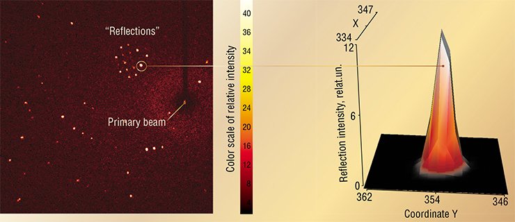
Investigation of the crystal structure is carried out using a complicated device, a single crystal diffractometer. The principle of its operation is based on capturing the diffraction patterns of X-rays passing through a single crystal. Very few laboratories can afford such an instrument, and it takes an experimental scientist with a great deal of practical experience to assess the reliability of crystal structure identification. If structural data are in doubt, the results of analysis based on them may be erroneous.
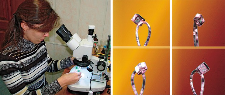
The work in this field is extremely varied and places great demands on the researcher. The search for a crystal suitable for the measurements is a creative job in itself, and not everyone succeeds in it. It is difficult to guess what should the shape of a “correct” crystal be, find it among many others and preserve until the measurements. Frequently, crystals will decompose, for example, due to amorphization in the air. So, all the selection work should be carried out relatively quickly. Auxiliary operations require skills comparable to the craftsmanship of Levsha shoeing a flea. It is necessary to place a tiny crystal with dimensions measuring fractions of a millimeter on a sample holder – a fragile glass wire or a nylon loop which is several times thinner than a human hair.
To choose the right strategy for the experiment, it is necessary to know well crystallography, the science mathematically describing possible types of symmetry occurring in crystals. Naturally, errors at any stage make it impossible to solve the structure of a new substance synthesized by chemists.
The experimental data obtained, amounting, after their mathematical processing, to some points in space, should be used to solve the structure of the crystal itself. The solution of the puzzle is complicated by several factors. First, some points related to experimental errors have to be recognized and discarded. Next, it is not known an atom of which element corresponds to each point. Finally, it is often impossible to predict what structure has to be found.
The success in solving depends on how well the researcher has learned to evaluate the effect of symmetry on the crystal structure and whether he/she is capable of recognizing structural fragments correctly. An unbiased and impartial attitude is a must because an expectation to see the desirable often impedes finding the answer. This is why each solution of a crystal structure is a new challenge to the researcher, which should be met with perseverance and sometimes even stubbornness.
X-ray structural analysis is now at its heyday and contributes to the development of other branches of chemistry by providing them with unique information on the composition and structure of just synthesized compounds. In turn, each structure solved gives new fundamental data for crystal chemistry itself, bringing us closer to the understanding of crystal.
The earliest diagnosis
Breast cancer is the most frequent type of cancer in women: more than a million women are diagnozed with it annually, and this figure is constantly growing. Hence, the death rate is constantly growing too, although with the help of modern methods of treatment almost a 100 percent survival rate has been achieved at the first stage of the disease. However, early tumor diagnosis remains a serious problem: a correct diagnosis is established in no more than half the cases. More than 80 % of patients discover the tumor themselves, by accident, when it has grown considerably.
Modern methods of breast cancer diagnosis are divided into two categories: instrumental (mammography, USI and tomography) and laboratory. In West European countries, mammography is usually used to screen for breast cancer and diagnoze women older than 35 years. The accuracy of tests reaches 80—98 %. Despite its high efficiency, this method is not recommended for examining young women and monitoring tumor growth. Women under the age of 35 should be examined using USI; however this method does not allow visualizing all forms of malignant tumors in breasts. In addition, sometimes areas of adipose tissue degeneration are mistaken for pathological structures.
Laboratory immunochemical and biochemical methods can only be used if the tumor has been formed and secretes enough of corresponding proteins-antigens in blood. That is why these methods are usually used to monitor the disease and evaluate the efficiency of antitumor therapy. Their diagnostic efficiency is lowered by the fact that protein markers are only detected in one third of cancer patients. However, a small number of them is present in healthy organisms as well as those with non-cancerous diseases.
Since the methods of early detection of mammary neoplasms are imperfect, completely new approaches based on the analysis of extracellular DNA and RNA are being developed.
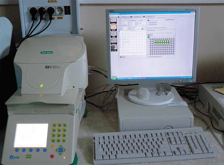
Until recently, it was considered that in higher organisms DNA is localized exclusively in cellular structures, namely, in cell nuclei and partially in mitochondria. In recent years though, it has been established that outside the cell, first of all in blood plasma, extracellular nucleic acids are found in both healthy organisms and those with pathologies. Their source is the cells in different tissues in the organism, including the neoplastic ones, which die as a result of apoptosis (cellular “suicide”) and necrosis.
According to Svetlana Tamkovich, it was the Academician V. V. Vlasov’s lecture in the course “Hot spots in molecular biology” which inspired her to start research in extracellular nucleic acids. Her graduation paper completed under the supervision of Candidate of Biology P. P. Laktionov was devoted to the development of adequate approaches to measuring the concentrations of nucleic acids circulating in blood and researching them in different pathologies.
Until now, neither the mechanisms responsible for the appearance and circulation of extracellular nucleic acids nor their biological functions have been studied sufficiently. Some data show that they can be actively secreted by the cells, with the frequency of some of these nucleotide sequences being different from that in genome DNA (Stroun et al., 2009). Besides, in the structure of circulating DNA, virus and bacteria DNA are detected as well as that of phytogeneous origin and DNA of the fetus which appears in the blood flow already during the first month of pregnancy (Tamkovich et al., 2008).
At present, attempts to create methods of cancer diagnostics on the basis of extracellular DNA are being made in many developed countries. A reason for this is that tumor specific DNA can be detected in the blood of cancer patients when a neoplasm exceeds 1 mm in size, i.e., when it consists of several hundreds of cells. The type of tumor and its localization do not matter in this case.
It is important that the analysis of changes in the composition and the amount of DNA circulating in blood can be used for the early detection of malignant tumors of any localization without a histological test. The same method can be used to evaluate the effectiveness of anticancer therapy. But it is not easy to track changes in the pool of extracellular DNA, which attest to the presence of a tumor process, using free extracellular nucleic acids from plasma or blood serum only. The effectiveness of such diagnosis is rather low and ranges from 10 to 50 %.
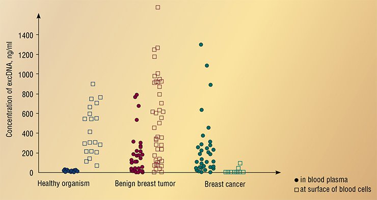
S. N. Tamkovich and her colleagues from the cell biology research group at the Institute of Chemical Biology and Fundamental Medicine SB RAS (Novosibirsk) were able to prove that in a healthy organism the overwhelming amount of extracellular DNA in blood is adsorbed on the surface of blood cells (Tamkovich et al., 2005). Circulating DNA and RNA enter the blood corpuscle due to ion interactions and through their connection to specific cell surface receptors. Using a total of extracellular DNA pool – both free and cell-surface-bound – for diagnosis makes it possible to detect cancer at the first stage in 95 % of cases, and also to differentiate the patients with benign tumors.
This is the approach to diagnosing malignant tumors that was used by the specialists from the cell biology research group ICBFM (SB RAS, Novosibirsk) when developing a new diagnostic platform for breast cancer. The ideological basis for the project was laid in 2004 and it was then that it was demonstrated that there is a principle possibility to carry it out if an appropriate technological base is available.
In comparison with the widely used methods of testing for oncomarkers, the new diagnostic platform has a number of important advantages.
First, traditional genetic diagnostic methods useful in cancer diagnosis are based on the detection of malignant gene mutations. However, the frequency of such mutations can vary widely depending on the type of the tumor. That is why even if a broad set of marked mutations is used, it cannot recognize the process of oncotransfomation unmistakably.
For early oncodiagnosing as molecular genetic markers of circulating DNA they use the so-called epigenetic markers, which allow detecting hypermethylation of promoter (regulatory) areas of a number of genes. The latter are the genes which participate in regulating the cellular cycle and setting off apoptosis, cellular differentiation, DNA reparation, etc. As a result of complete deactivation of regulatory areas even when there are no structural changes in the genes, they become inactive, hence a cell can become neoplastic. That is why thanks to epigenetic markers the precision of diagnosing can improve considerably.
Second, for effective extracellular DNA separation and purification from blood scientists from Siberia use their own original technologies which are protected by six Russian and one European patents.
At present the researchers are working on optimizing the stages of analysis without lowering the method’s validity and sensitivity to meet the modern standards of clinical laboratory diagnostics. It requires making the procedure of blood processing simpler and using a new method of detecting marker genetic sequences (multiplex PCR-analysis in real time).
To prove the diagnostic significance of the new method, an extensive information database and a collection of blood samples from the patients with established diagnosis as well as from new patients with suspicion of tumor are being created. This work is being done in cooperation with Novosibirsk Regional Cancer Hospital, Tsentralny District Maternity center and Central Clinical Hospital in Sovetsky district of Novosibirsk.
For the future of the new method of breast cancer diagnosis, it is quite important that it is noninvasive (i. e., it does not require making biopsy for the histological test) and it does not require expensive equipment. It means that it can be widely used to assess the effectiveness of antitumor therapy and in screening programs aimed at detecting this widely spread oncological disease.


