"...Almost Immortal and Forever Young"
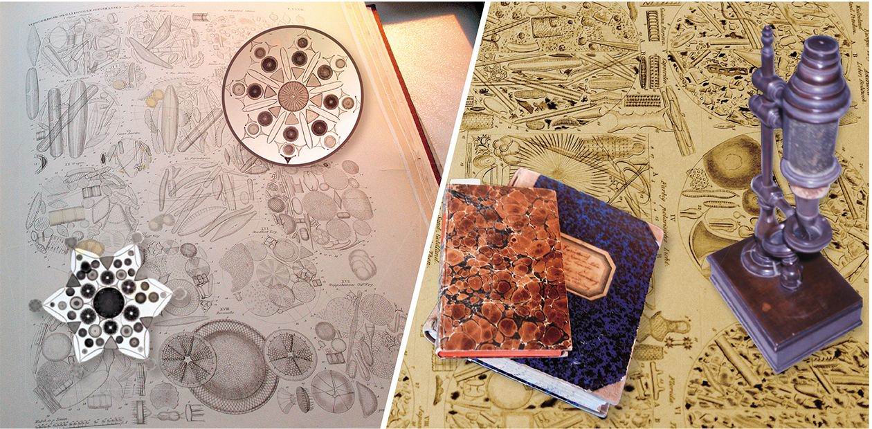
Because of their extreme minuteness and the transparency of the glass wall of the diatoms, their discovery and existence was only made possible by the invention of the microscope — in the 17th century. The structure of the walls was used in the 1700 and 1800s to assess the strength of lenses made for microscopes. As improvements were made to the microscope new details of diatom wall structure were observed…
230 million years under water
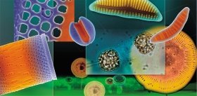
It is often said that the diatoms live inside a glass house. In fact, their hallmark is their unique type of cell wall composed of silica, which consists of two parts that fit together like a pill box with a series of bands to hold pieces together. Through the holes in the wall the living cell can communicate with the outside world and get its food, oxygen and carbon dioxide for photosynthesis.
Scientists who study diatoms boil the living cells in acid to remove all the cell contents and make the pill box-shaped cell walls with their bands come apart. The empty cell walls are then mounted in a refractive fluid that makes the ornamentation of the cell wall stand out from the background around them. The ornamentation on the walls is used to identify the kind of diatom it is. Depending on how strong a microscope you have, more or less detail can be seen on the cell wall.
Because the diatoms have a glass cell wall, the glass wall is left behind if and when they die, then. The cell wall falls to the bottom of the place where they lived and is incorporated into the sediments. This has happened for millions of years and the diatoms have a long fossil record. Diatom glass cell walls often preserve well, with their intricate microscopic details largely intact, and can be identifiable to species level after millions of years.
Because of their extreme minuteness and the transparency of the glass wall of the diatoms, their discovery and existence was only made possible by the invention of the microscope — in the 17th century. The structure of the walls was used in the 1700 and 1800s to assess the strength of lenses made for microscopes. As improvements were made to the microscope new details of diatom wall structure were observed.
The manufacture of microscopes in 19th century in Germany, Great Britain, France and America led to their presence in most middle-class Victorian homes. Most microscopists at this period were interested in many microscopic organisms, such as diatoms, dinoflagellates, radiolarians and protozoa. Prepared slides of diatoms in all their elegance were an important acquisition and facilitated exchange of information about diatom structures between amateur enthusiasts in microscopical societies, which were prominent in Victorian times. These societies were set up in most of the larger cities and their periodicals published results of these early amateur diatomists.
Slides of diatom material were exchanged and individual diatom cells were marked on the slide encircled by a diamond marker that scratched on the surface of the cover glass. Diatomists sold exsiccatae sets of dried material with labels of identification and locality information. A number of enthusiasts made their living by selecting diatoms individually and placing them into rows or patterns, providing list of names to go with the arrangements and then selling them.
The diatoms were selected individually by hand using a pig’s eyelash or a micro-manipulator. The selected cell was then moved to a cover glass onto which a minute drop of gum tragacanth had been dried. They could then be arranged into rows according to genera, locality, etc. or into intricate patterns. The arrangement was fixed by breathing onto the cover glass and the gum tragacanth kept the cells in position
Molecular clocks have been made to estimate their time of origin. These calculations show that the diatoms probably evolved about 230 million years ago. These first diatoms probably were not living in the glass houses that they live in now. Instead, they were likely to have been naked flagellated cells.
The very first diatom deposit has been found in Korea from a terrestrial deposit. How can it be that the diatoms first lived on land? It has been hypothesized that when the oceans flooded the land 230 million years ago, pools of water were left behind with the early flagellated diatoms in them. The diatoms began to use silica to put the cells into a resting state in these small ponds because silica can be used for chemical reactions that keep the cell from ageing.
They took up so much silica that the silica precipitated because it was in too high a concentration. The first precipitated silica was likely in the form of small scales, such as you see today in the wall of the modern zygote. These scales were pushed to the outside of the cell, which helped to protect the cell from drying out in the small ponds. Thus, the naked cell developed a need to bring silica into the cell to keep it from ageing and then it precipitated the silica to form a protective layer to keep it from drying out. Eventually two of the scales were changed into the two parts of the pill box and the remaining scales were changed into the bands holding the pill-box together.
For descriptive purposes diatoms fall into two broad groups, based on the organisation of the pattern of ribs and pores on the valve face, centric diatoms and pennate diatoms. The relationships among the diatoms have been investigated by analyzing their genome. These results show that there are not two groups of diatoms as you would think from the pattern on their cell walls but instead there are three groups: two centric groups and one pennate group. Each group has a different type of sexual reproduction.
Eventually, the size becomes so small that the cells die. Evolution has been kind to the diatoms and they have invented a way to become large again. They undergo sexual reproduction. The cells throw off their walls, become a gamete (male or female), find a mate and fuse to make a zygote or fertilised cell. The naked zygote swells to become larger and larger. It can swell because it has a large central vacuole that fills and fills with water. When large enough, the cells then reform their silica cell wall or pill box and the cycle starts over again with one of the cells smaller than the parent cell each time they divide
The earliest evolved diatom in the molecular trees is a marine representative of a terrestrial diatom, Ellerbeckia, which would lend support to this hypothesis of where the diatoms may have evolved. The next fossil record of the diatoms is in the early Cretaceous and these are totally marine diatoms. Thus, it would seem that the early terrestrial diatoms were washed back into the sea the next time that the oceans flooded the land. These diatoms in the early Cretaceous are cells with very unusual, thick and ornate cell walls with strong mechanisms to link the cells into chains. They look very different from modern diatoms. These diatoms are so heavy that it is likely that they lived in near-shore marine, turbulent environments where they were semi-benthic or epiphytic or attached to rocks.
Deposits of diatoms continue through geological time to the modern day. Diatoms occur in all major types of waters and on sediments or solid surfaces that are kept wet. It would be fair to say that every other breath of oxygen that we breathe comes from diatoms.
They are nearly all photosynthetic and make their own food. As primary producers in the world’s oceans, they are major players in the global cycling of nutrients and form the base of the food chain in aquatic ecosystems for the zooplankton to eat. Just a few hundred species in the marine phytoplankton fix around 20 % of total carbon to be used as food for the animals in the ocean.
The complete genome has been sequenced from two diatoms. Both of them show that the diatoms are half animal and half plant. This is a reflection of their evolutionary history because millions of years ago a flagellated animal like unicell engulfed a unicellular alga and stole its plastids and kept them for themselves so that they would always have a food supply. This is called endosymbiosis and that’s why part of the gene from the diatom belongs to the animal host and part belongs to the alga it engulfed. The diatoms can make urea just like animals do, only they don’t need to urinate instead they use urea as a starting products for other chemical reactions in the cell.
We don’t fully understand why the diatoms make urea. In fact, we can only identify the use of about 60 % of the genes in their genome. What novel products do the unknown genes make? This is why genome sequencing is so exciting because not only do you find the genes that you know about but also you find many, many genes with an unknown function.
R. CRAWFORD: These deposits from the early Cenozoic, over 35 mya, even the Mezozoic, over 65 mya were magnificent in their multitude of experimental forms, hugely robust and deposited in such numbers that we can only wonder at what the seas looked like. They were probably turbulent and very productive over shallow shelves that must have fed the deposited remains of the dead cells to greater depths to be concentrated in such numbers that they can be mined and quarried today.
Some species have been kept safe and flourish now, tens of million years after their first appearance, and, moreover, have their doubles in modern communities.
In some extinct, fossil diatoms whose history goes back as far as the Cretaceous (over 65 million years ago), we find massive, and sometimes elaborate structures at the centre of the valve face. In others the linkage is found to one side or all over the valve surface or it may be found around the valve rim only. The design of these spines is often more than mechanism constructed to keep cells from drifting apart and this prompted us to examine the forces acting on the cells in a turbulent water.
The chloroplasts of the penetration group: the history of one discovery
Russian scientists have greatly contributed to the investigation of diatoms at all stages. Especially important are works by K. S. Merezhkovsky (1855—1921) and, specifically, his most essential general biological conclusions about the evolutional role of symbiosis and origin of organisms.
K. S. Merezhkovsky’s basic theoretical views in this field formed during the study of the chloroplasts of diatoms where photosynthesis takes place. “In no other group of plants do chromatophores play such a great morphological role and, therefore, acquire such great importance and are of such interest as in the diatom class… As if a peculiar, unique, and fascinating world of phenomena opens up for us; inside the diatom cell we come across some self-maintained organisms that are independent of it, live in it as “guests”, develop, divide, and propagate. They don’t destroy the cell itself, and depend on it only to the extent to which organisms in general depend on the environment. There is nothing of the kind, at least in such immense sizes, in the plant kingdom” (Merezhkovsky, 1906, “Endochrome Laws”, p. 5).
The pioneering article by K. S. Merezhkovsky “On the Nature and Origin of Chromatophores in the Plant Kingdom”, which appeared in 1905, made his name famous in the history of science. In this article, he clearly formulated the hypothesis that chloroplasts are photosynthesizing organisms-symbionts, which intrude into the cell and become there major suppliers of nutrients. The term “symbiogenesis” was also offered by him.
Millions of years ago a flagellated animal-like unicell engulfed an alga, stole its plastids and kept them for themselves so that they would always have a food supply
In 1909 Merezhkovsky published the article “Theory of Two Plasmas as a Basis of Symbiogenesis, a New Theory of the Origin of Organisms”. In this article he writes, “If there exist two plasmas of fundamentally different properties and, hence, two worlds of organic substances, this can be explained only by the fact that both of these plasmas originated independently, in absolutely different conditions and, hence, in different epochs of the Earth’s history” (K. S. Merezhkovsky, 1909, p. 87). It should be noted that a French researcher E. Shatton suggested dividing the entire organic world into two basic groups — procaryotes and eukaryotes 16 years later! One can only wonder at K. S. Merezhkovsky’s striking scientific vision and be thankful that it was the study of diatoms that brought him to these outstanding discoveries.
Generally the history of any science consists of stages determined by major discoveries and achievements of a certain period. The latter are ultimately due to the level of development of the society and technical capabilities. However, people have always been the main “driving force” of the scientific process. These are personalities attracted by the Unknown, which makes them permanently seek answers to the questions “why” and ‘how”.
It is a hard task to write the history of diatomology. The difficulties are mainly due to the fact that too few materials about those scientists who contributed to our modern knowledge of the world of diatoms remained. In a short article, it is impossible even to list the names of all researchers, because it would then become a reference book. The creation of a comprehensive history of diatomology with detailed data about the researchers should become one of the tasks of the International Society of Diatomologists, under whose aegis International Diatom Symposia are held in different countries once every two years.
Siberia under microscope
When Ehrenberg (1795—1876) was invited by Alexander von Humboldt to accompany him in 1829 on his journey to Russia he was already known as a promising young biologist who had collected and described many animals and plants from Northern Africa and the Near East. Ehrenberg’s favourite subject of inquiry was microscopical organisms, called then “Infusionsthierchen”
Ehrenberg writes afterwards about this Russian trip: “Mr. Baron Alexander von Humboldt’s journey to the Russian provinces up into the Chinese “Dzungarei” [today known as the northern part of the Chinese Province Xinjiang] to which I was honorably and cordially invited and which… gave me the opportunity to get to know a decisive part of the surface of the earth… I have not refrained from putting my ceaseless attention on the geographical relation of the smallest forms of organic life in such big parts of land. These new and numerous observations… made me discover a great deal of unknown forms and gave me a survey of the geographical relation…”
Christian Gotfried EHRENBERG (1795—1876) was a German naturalist-zoologist, well known by his works on morphology, physiology and taxonomy of microorganisms. He had in his possession one of the largest collections of his time and was called “Mr.Microscope” by his contemporaries. He was the foreign corresponding member and honorary member of the Saint Petersburg Academy of Sciences
Humboldt had been invited by Czar Nikolaus 1st to investigate the natural and mineral resources of the Ural and West Siberia. The expedition began in Berlin on 12 April 1829 and continued almost nine months. All along the tour they were met and accompanied by Russian fellow scientists.
Humboldt’s route of the expedition is well recorded but Ehrenberg’s point of view is a little different as he writes mainly about the waterbodies they met and in which he took samples: at the River Neva, the River Volga, Ural, Irtysh, Obey… And in every drop of water he met with the world of miraculous creatures again and again. In his Siberia paper published right after his trip, Ehrenberg defined infusoria: “Infusoria including the smallest flagellates are not slime without structure, but they are organized at least with a mouth and digestive system.” (1830). Ehrenberg’s infusoria included all microscopical organisms from the bacteria, colorless and colored flagellates, to diatoms and even rotatoria. Just as with visible animals, he saw in them complete organisms endowed with organs. This was against the trend of his time which saw in them structureless forms which appeared out of nothing.
Ehrenberg used the microscope routinely; he even carried a microscope with him on all his journeys, taking samples in the daytime and drawing the organisms at night. On his travels, Ehrenberg drew on small pieces of paper, adding a hand written manuscript name, sampling place and size. Later, the small drawings were glued on standard paper sheets, which were inventoried by Ehrenberg’s daughter Clara. Ehrenberg wrote that he was able to measure dead or not moving infusoria down to 1/4000 of a Paris line (1 Paris line = 2256 µm; 1/4000 µm = 0.6 µm). Ehrenberg’s materials and preparations as well as his drawings and letters survived World War II and are today stored in the Ehrenberg Collection in Berlin (Humboldt Universitt zu Berlin). Recently they were digitized and databased by the curator Dr. David Lazarus and co-workers
Ehrenberg was eager to prove to the world that his microscopical findings were correct and could be re-investigated. Whenever possible he took samples back to Berlin. After his journey to Siberia he began to build up a collection of preparations of infusoria, mainly diatoms which attracted him for the rest of his lifetime because their “valves build up indestructible soils, stones and rocks”.
Ehrenberg cultivated living cells for further studies in vivo. He isolated single cells with a pipette and put them into a test-tube. In this way, he collected “a graceful menagerie of living infusoria” and stored his living collection on the window sill in order to enlarge the time span of studies and to over winter living cells.
Ehrenberg had proven time and again that no infusoria would appear de novo. ”
…The studied reproduction of infusoria by self-division ... shows their sustainability and distribution in oceans and winds which poetically come close to immortality and eternal youth. One may divide oneself into countless new parts in order to stay forever young for countless years.” (1838)
C. G. Ehrenberg: “I am ending this communication with the feeling that I was not able to fathom this profundity of organismic creation. To have opened it should be my excuse for time and energy spent. May fresh and soulful eyes delve into nature and collect which it conceals — not purposeless — in darkness and smallness” (1830)
Ehrenberg wanted to resolve if infusoria are commonly distributed, that means if there are cosmopolitans, “world citizens”, among them. His conclusion drawn from his journey to Sibiria can be read in his book “Infusionsthierchen”: “the existence of infusoria has been proven in all four continents and in the ocean and some species are the same in the most remote areas of the world … The geographical distribution of the infusoria on the earth follows the laws which have been recognized already with other natural bodies: towards the south the taxa are more different than towards the east or west.” (1838).
Darwin and diatoms
Darwin began the Origin of Species with these words: “When on board H.M.S. Beagle, as naturalist, I was much struck with certain facts in the distribution of the inhabitants of South America, and in the geological relations of the present to the past inhabitants of that continent. These facts seemed to me to throw some light on the origin of species — that mystery of mysteries, as it has been called by one of our greatest philosophers.” (1859).
As inspiration, Darwin suggests two factors: biogeography – “the distribution of the inhabitants of South America…” – and history – “…the geological relations of the present to the past inhabitants of that continent”.
Darwin made some comments on diatoms and their distribution in the first edition of his Journal of the Voyage of the Beagle (1839). He made some remarks on Christian Gottfried Ehrenberg’s study of the face paint used by the local people in Tierra del Fuego: “This substance, when dry, is tolerably compact, and of little specific gravity: Professor Ehrenberg has examined it: he states…that it is composed of infusoria [which included diatoms]… He says that they are all inhabitants of fresh-water; this is a beautiful example of the results obtainable through Professor Ehrenberg’s microscopic researches…”
Darwin further notes: “It is, moreover, a striking fact that in the geographical distribution of the infusoria, which are well known to have very wide ranges, that all the species in this substance, although brought from the extreme southern point of Tierra del Fuego, are old, known forms”.
Darwin suggested that diatoms have very wide geographical ranges and because these are old known forms, diatom distribution patterns are old. Darwin’s assumption that they were known forms is correct – but they were known and described by Ehrenberg from a restricted part of the world. In other words, Ehrenberg had seen the usual kind of mix one encounters in any flora, diatom or otherwise – some species are widespread, some not so. Ehrenberg made some general comments on distribution, first published in 1849: “... the Rocky Mountains are a more powerful barrier between the two sides of America, than the Pacific Ocean between America and China; the infusorial forms of Oregon and California being wholly different from those of the east side of the mountains, while they are partly identical with Siberian species.”
Ehrenberg understood that a relationship of some kind existed between the diatoms from both coasts of the Pacific and thus states the first cogent reason for studying diatom distribution patterns.


