The Fungal Lilliput: From Parasites to Predators
Gathering robust ceps in the fall, we do not keep in mind that they are not the fungi themselves but only their fruiting bodies, although quite large. From the systematic standpoint, all the fungi belong to microorganisms; indeed, the overwhelming majority of fungi are visible with the naked eye only when they form colonies growing on an appropriate substrate. This large invisible microcosm is all around us. The fungi are everywhere: in the air and in the soil, in houses and in museum pieces, and even in our morning sandwich…
Among higher organisms fungi form a vast separate kingdom*. Taxonomists consider two divisions of this kingdom, namely, the slime molds Myxomycota and the true fungi Eumycota, which, in turn, are subdivided into six classes. Amateurs can hardly grasp all these details of fungal diversity and, as a matter of fact, they do not need them. Moreover, mycologists themselves, for convenience, divide all the fungi into two groups – micromycetes and macromycetes – independently of their taxonomic positions.
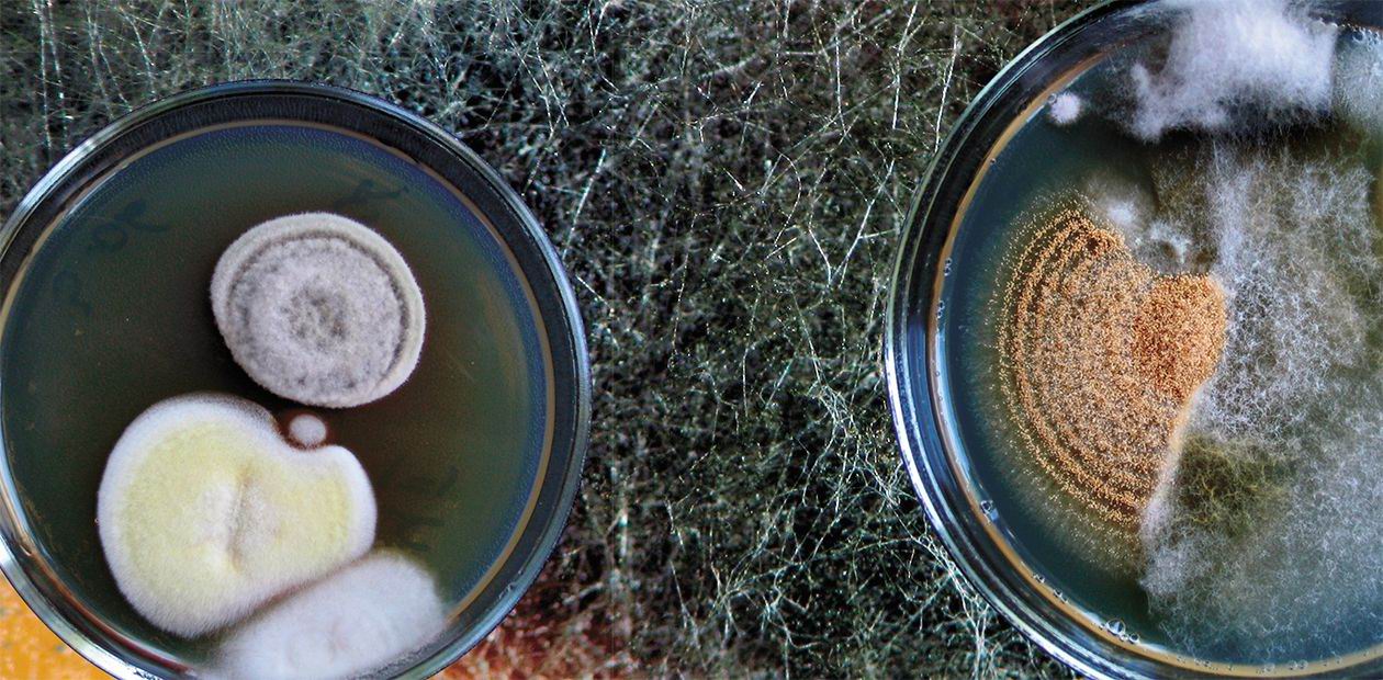
Micromycetes (micros is Greek for “small” and mykes is Greek for “fungus”) justify their name as visible only with the help of a microscope. However, when growing on an appropriate substrate, micromycetes become visible with the naked eye, forming the commonly known “molds.” Nonetheless, they never develop large fruiting bodies as, for example, pileate fungi, widely used in cooking and medicine.
However, people have long ago learned to use these subtle and frequently rather unattractive creatures to their advantage. Wine production and bread baking are examples of the first biotechnologies that utilized yeasts, the unicellular representatives of a vast class of cup fungi (morels and famous truffles belong here). The fungi of the genus Penicillium, which gives a sharp taste and a blue marble coloration to the famous Roquefort, have gone down in the history of cheese making. The same genus opened a new era in treating severe bacterial infections, having granted to the world antibiotics.
Initially, people used wild-type strains, but later selection and advances in genetics allowed highly efficient micromycete strains to be obtained; these strains are now widely used not only for producing wines, cheese, and antibiotics, but also for synthesizing various organic acids, enzymes, vitamins, food supplements, and so on. From the very beginning, development of biological tools for plant protection against insect pests, diseases, and weeds has involved microscopic fungi.
However, each coin has two sides – many species of micromycetes can cause various plant, animal, and human diseases. The presence of spores of the fungi belonging to the genera Aspergillus, Cladosporium, and Penicillium is a risk factor for human health, since some of its representatives can cause serious diseases (mycoses) or allergies (Ivanova et al., 2007; Kononenko et al., 2008). Pathogenic and allergenic species of microscopic fungi are isolated even from the museum air and exhibition pieces (Bogomolov et al., 2007).
To sum up, the microscopic fungi, both useful and harmful for humans, are immediately near us, inhabiting soil, air, walls of our residences, and so on. These microorganisms, as has been demonstrated, are integral components of any terrestrial ecosystems.
Just out of the air
To make sure that micromycetes are ubiquitous, it is enough to look through the data of biological monitoring of the atmospheric aerosol in the south of West Siberia obtained by several scientific departments of the of the SRC VB Vector (Safatov, Teplyakova, et al., 2009).
The fungi detected in the cultivated atmospheric air samples contained representatives of 18 genera excluding unidentified specimens. The fungi belonging to the genera Aspergillus, Penicillium, Cladosporium, and Alternaria were the most abundant; all these fungal species, potentially harmful for human health, are also characteristic of other CIS regions, from Adjaria to St. Petersburg (Ivanova et al., 2007; Verulidze et al., 2008). The representatives of another five species are phytopathogenic, i. e., they cause plant diseases.
However, the cultivated atmospheric air samples also contained colonies of the fungi that were potential producers of various biologically active substances, for example, the dark-colored fungi containing melanin, whose spores were present in all the atmospheric air samples.
As is known, melanin (melanos is Greek for “black”) is a black pigment widespread in nature, contained in the epidermis, hair, retina, etc.; large amounts of this particular pigment are formed in our skin exposed to ultraviolet. However, its functions are not confined to providing sun tan: melanin is not only a regulator of cell metabolism, but also plays the role of the universal protector in the cells exposed to various physicochemical factors of mutagenic and carcinogenic nature (Borshchevskaya et al., 1999).
Currently, ointments containing fungal melanin and intended for healing skin diseases are produced in Belarus (Litvinov et al., 2008). Fungal melanins are also studied in Irkutsk (Ogarkov et al., 2008).
Of special interest for medical biotechnology among the fungi found in the cultivated air samples is the genus Aureobasidium. Commercial production of a new plasma substitute using a species from this genus was launched in Belarus (Litvinov et al., 2008).
“Free-riders”
The fungal colonies grown from atmospheric air samples are frequently a mixture of two or more species, with individual colonies differing in their shape and color. A comprehensive microscopic examination demonstrated that frequently one fungal species parasitized on the other.
The term mycophily means the ability of an organism to develop in nature at the expense of fungi. Viruses, bacteria, actinomycetes, etc., can be mycophils. However, fungi exceed all these organisms in the number of species and their parasitic activity; this group is named correspondingly – the mycophilic fungi. Containing the representatives of almost all fungal classes, this group includes two thousand species (Rudakov, 1981).
Mycophilic fungi are abundant in various climatic zones and in all types of habitats, including water, soil, fruiting bodies and mycelia of macroscopic fungi, and the surface and inner parts of various micromycetes. These fungi play an important role in the natural ecosystems by enhancing degradation and mineralization of fungal remains and limiting the population sizes of other fungi.
Mycophilic fungi are natural enemies of phytopathogenic fungi; therefore, they are used for biological plant protection. For example, the fungi from the genera Trichoderma and Ampelomyces as well as some others are producers of the corresponding biological preparations. On the other hand, mycophils present a serious threat to the cultivated edible mushrooms; it is enough to mention only the wet bubble disease of champignons, caused by the fungus Mycogone perniciosa. The yield of champignons infected with this pathogen decreases to one half or less.
Parasitic fungi can also play a negative role in several other cases associated with fungi cultivation, in particular, in keeping the collections of fungal strains when colonies are reinoculated to fresh nutrient media, or in the production of commercial mycelia. Note that the development of commercial production of fungi in Russia has given rise to numerous laboratories involved in growing mycelial inocula. It is no secret that such laboratories frequently lack even a microscope and estimate the mycelium growth and quality only visually according to the colony color and growth rate.
The mycophilic fungi present a special threat when tissue and spore cultures are isolated from natural or cultivated fungi. For example, almost all strains of edible basidiomycetes selected in the 1950s—1970s for commercial cultivation to produce fungal biomass eventually appeared to be mycophilic hyphomycetes.
Correspondingly, cultivation of such strains gave birth to the erroneous concept that a submerged cultivation of basidiomycetes produced the mutants with the sporulation type untypical of higher fungi. Since the fungal biomass produced by such cultivation lacked the expected taste and aroma, this had a negative effect on further research into submerged cultivation of edible fungi (Bukhalo, 1998).
Our studies have demonstrated that mycophilic fungi can persist in the cultures of many edible and medicinal fungi as mycelia, i.e., the hyphae of a parasite are retained in the hyphae of a basidiomycete (Teplyakova, 1999). Moreover, their presence in the reinoculated colonies can be concealed for a long time, being detectable only by microscopy.
However, sometimes a parasitic fungus can get an advantage in development, and a mycologist unacquainted with specific features of mycophilic fungi may fail in puzzling out why another fungus, not the one that is cultivated, reproduces massively. The most frequent explanation is an insufficiently sterile medium; however, the actual reason is much deeper.
Eventually, a basidiomycete culture can be lost at all. For example, a patented champignon strain, the producer of a biologically active substance, was brought to Novosibirsk from Kazakhstan. The author of this strain, an experienced technologist, controlled the fungal culture after its isolation from nature mainly visually. As a result, it was found out that the producer of a biologically active substance had long been a mycophilic fungus from the genus Verticillium rather than the patented champignon.
Nematode hunters
Of special interest among micromycetes is one ecological group of fungi that is evolutionarily connected with nematodes (roundworms), common soil dwellers. These amazing fungi, which can be formally attributed to parasites, are actually genuine predators.
The natural enemies of nematodes are referred to as predatory fungi for their ability to develop various tools for trapping their prey. Predatory fungi have been discovered in almost every part of the world, which suggests that they play an important ecological role by utilizing a tremendous mass of nematodes, many of which are agents of dangerous plant and animal helminthoses.
In general, the role of predatory fungi in nature has not been studied sufficiently, despite considerable efforts made recently to isolate efficient strains of nematophagous fungi from nature, examine their specific features, develop technologies for producing biopreparations against various phytoparasitic nematodes, and test these preparations in nature (Teplyakova and Anan’ko, 2009a, b).
A specific feature of the fungi is a vast diversity of organs and reproduction systems. Fungi can reproduce vegetatively, asexually, and sexually. Since the appearance of a fungus can change strikingly with the change in the reproduction habit, it is not surprising that they can be taken for separate species.Vegetative reproduction requires no specialized organs; it is implemented via mere parts of the mycelium or individual cells formed by budding of the threadlike hyphae. Reproduction via chlamydospores, special thick-wall cells developed on the mycelium, also belongs to the vegetative type.
The marker of asexual and sexual reproduction types are specialized spore cells, which are analogs of the seeds produced by higher plants. The fungal spores are usually immotile; very small in some species, spores can be transported over tremendous distances and ascended to a high altitude. In some species, spores can be disseminated by insects or animals, while other fungi are able to launch spores as if with a catapult, shooting them into the air.
In an asexual reproduction, spores are formed on specialized hyphae of the aerial mycelium. The spores formed at the top of hyphae are named conidia and these hyphae, conidiophores.
In addition, spores can form endogenously, inside the specialized cells at the end of conidiophores (sporangiospores).
In a sexual reproduction of fungi, the sexual process – fusion of gametes followed by pooling of the genetic material from their nuclei – precedes spore formation. The process takes place in specialized reproductive organs, for example, the so-called sacs (ascospores in morels and ergot fungi) or in basidia (basidiospores in many forest mushrooms)
However, justice was found for these minute predators, as well, and again, these were mycophilic fungi. Researchers from the SRC VB Vector encountered this phenomenon when isolating hyphomycetes from soil and cultivating them: a detailed microscopic examination has demonstrated that hyphae and conidia of a predatory fungus sometimes appear completely deprived of their “living” contents due to the parasitizing of a mycophilic fungus. Although the growth of a parasite in the colony of a predator could have been unnoticed, this frequently led to the replacement of the host. Consequently, the quality of a biopreparation against nematodes decreased, eliminating the expected effect of its application to soil.
On the other hand, there are opposite examples. In particular, a Siberian strain of the predatory fungus Arthrobotrys longa was studied at the Laboratory of Antibiotics of Moscow State University as a potential producer of fibrinolytic enzymes. However, a careful comprehensive study of the strain demonstrated that the producer of these enzymes was an accompanying mycophilic fungus from the genus Cephalosporium, rather than the predatory fungus.
Further assessment of three isolates of mycophilic fungi recovered from predatory fungi confirmed the assumption that these parasites were the true producers of fibrinolytic enzymes (Teplyakova, 1999). Perhaps, it is this group of mycophilic fungi that should be searched for the fungal strains with a fibrinolytic activity.
Among the higher organisms, fungi are the champions in the ability to adapt to most diverse environmental conditions. The developed surface of their threadlike hyphae, forming the fungal mycelium, provides a large area for absorption-based uptake of nutrients. Possessing powerful enzyme machinery, fungi are able to destruct many man-made materials, including wooden constructions, building materials, and even aviation fuel.
Taking into account the danger of biological damages, the construction regulations since 1997 contain the term “biologically active media” and scientists propose the regular technical surveys of aircrafts should include obligatory control of the fuel and fuel systems by standard mycological and microbiological tests.
However, as it has been repeatedly mentioned above, our long-standing enemies can become our allies, for instance, in curing human maladies. Indeed, fungi, including their microscopic species, are rarely used for pharmacological purposes; therefore, a challenge for scientists is to continue the search for efficient producers of biologically active substances among the tremendous natural diversity.
References
Borshchevskaya, M. I. and Vasil’eva, S. M. Development of the Concepts on Biochemistry and Pharmacology of Melanin Pigments // Vopr. Med. Khimii, 1999. V. 45. No. 1. P. 13—23.
Ogarkov, B. N., Ogarkova, G. R., and Samusenok, L. V. Fungi: Protectors, Healers, and Destructors. Irkutsk: GU NTs RVKh VSNTs SO RAMN, 2008.
Rudakov, O. L. Mycophilic Fungi, Their Biology, and Practical Importance. M.: Nauka, 1981.
Safatov, A. S., Teplyakova, T. V., Belan, B. D., et al. Concentration and Variation of the Mycomycete Composition in the Atmospheric Aerosol in the South of West Siberia // Optika Atmosf. Okeana, 2009. V. 22. No. 9. P. 901—907.
Teplyakova, T. V. Bioecological Aspects in Studying and Applying Predatory Hyphomycetes. Novosibirsk, 1999.
Teplyakova, T. V. and Anan’ko, G. G. RF Patent no. 2366178. A Method for Producing a Preparation Based on Chlamydospores of a Microscopic Fungus for Controlling Parasitic Nematodes of Plants and Animals // Byull., 2009. No. 25.
Teplyakova, T. V. and Anan’ko, G. G. Predatory Hyphomycetes against Parasitic Nematodes // Zashchita Karantin Rast., 2009. No. 6. P. 22—25.
Photos are courtesy of the author
* SCIENCE First Hand (Russ.), 2010. No. 33 (3). P. 104—113


