The Postgenomic Era: “Seeing through a Brick Wall”
Many people believe that experiments with animals always involve vivisection, although it may be anesthetized. This is in no way true. Many methods devised to date allow studying animals, various forms of their behavior, as well as their physiological and biochemical processes without any noticeable damage to their life and even health
Many people believe that experiments with animals always involve vivisection, although it may be anesthetized. This is in no way true. Many methods devised to date allow studying animals, various forms of their behavior, as well as their physiological and biochemical processes without any noticeable damage to their life and even health. This advance is connected not only with a graduate refinement of ethics in the human society, but rather with pragmatic considerations: scientific data obtained in the experiments with untraumatized healthy animals are less affected by experimental artifacts. Many of such methods have a long history, and their present advance is mainly attributed to providing comfortable conditions for both the experimental animal and experimenter himself/herself. Along with the improvement of traditional methods, an impressive success has been recently achieved in developing basically new approaches to imaging the inner structures and processes.
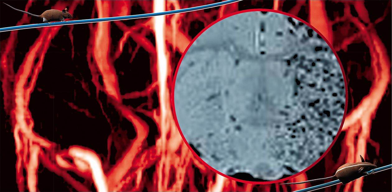
Tomography is a leading method in this field; this technology makes it possible to image internal organs by sections and reconstruct their 3D structure with the help of a computer. Depending on the physical principles underlying specific devices, they “see” various tissues of the body in different ways. For example, the brain tissues contain considerable amount of water (here, Aristotle was right) and, consequently, hydrogen atoms; therefore, it is better to scan the brain with the help of an NMR tomograph. The mice, which are rather small, need specialized “mouse-scale” devices. And such devices have been designed. Engineers succeeded in increasing the resolution of NMR tomographs to several tens of microns (!), which is a good accuracy even given the size of mouse organs. Such devices are very expensive – they are a luxury no academic lab involved in basic research can afford. However, a shared access center can bear the expense of this equipment and can thus make such studies affordable for many labs. This demonstrates, once again, that expensive experiments should be provided with high quality standardized animals, which will allow obtaining reliable results with a small number of replicates.
Why should we go to such great expenses? To get the opportunity to gain an ever deeper insight into the living processes that occur in the body. One of the most advanced models of such devices, Bruker Bio Spin, is planned to be acquired for the Shared Access Center for Genetic Models; this device is able to image moving tissues such as the blood and heart muscle, and gives challenging opportunities for experiments with a live untraumatized mouse. The same device or, more precisely, a system of devices, is able to supplement the data obtained in a new field of radiomedicine regarded as promising—the proton emission tomography (PRT). A state-of-the-art research tomograph has another extremely attractive function—its ability to spectroscopically assay the substance at a specified site of the living untraumatized organism.
The methods for optical examination of the body have also advanced considerably. The trend is just the same—search for the methods that will allow us both to examine animals and preserve their lives and, if possible, comfort. The light-sensitive devices capable of recording individual photons can visualize the luminescing tracer molecules directly through mouse tissues; however, the mouse should be at least white or, much better, bald (there are such animals). The current research into tumor growth and metastasizing utilizes cells labeled with genes producing a luminescence protein, luciferase. It is possible to optically observe the proteins that do not generate light themselves and yet can do it when excited by external radiation. Such induced light provides for visualizing the molecular structures, which irradiate it even through the body tissues. Equipment making it possible to induce and record radiation of native molecules in cells or intercellular space, deep in tissues (of special interest is the brain) is now considered promising and is being developed in many leading medical universities of the world. The traditional X-ray examination has acquired novel functions. Devices capable of analyzing the spectral composition the X rays that pass through the body have been designed; this allows the compositions of bone, adipose, and other tissues to be studied, which is indispensable for models of some diseases connected with the abnormalities in mineral and fat metabolisms. Concurrently with the radiation-based examination techniques, traditional invasive methods were developed. However, few of them are bloody as interventions with the body are minute.
Solutions of many applied and basic problems demand that researchers know concentrations of particular substances in a studied tissue or, on the contrary, inject specified amounts of preparations to certain organs. Moreover, it should be done so that the animal does not notice the invasion and, of course, it should be painless. Traditionally, this has been done via biopsies, at best – procedures often performed using anesthesia. Today, there is a range of microdialysis devices that can stay inside the body constantly and painlessly to sample a tissue fluid or apply a tested substance unnoticeably to the animal.
Let me complete this summary on some state-of-the-art methods used in biomedical research on a philosophical note. All methods, even the best, have their shortcomings. The advantage of the above mentioned methods is their efficiency, whereas the other side of the coin is their high cost and complexity in use. Studies involving such methods require considerable funding for equipment, training of the staff, and salaries. However, the results pay their way. This reminds of the Biblical story about talents saying that the richer gains more…


