The Postgenomic Era, or Why We Need 300,000 Mice Strains
There is no need to prove that the question on how a living organism functions is topical for each of us. The only problem is to choose an adequate “tool” for knowledge acquisition. As a rule, research into any biological system be it a cell, individual, or biocenosis commences from a “morphological” structural description. However, though this stage is necessary, it alone is insufficient for comprehending the functioning mechanisms of a studied system.
A brilliant illustration of how extremely erroneous can be speculations based on only structure descriptions is the concepts of ancient scientists about the brain function. Aristotle, as well as his colleagues, regarded this “materials substrate” of the cognitive functions as the depository for water and mucus (serving to cool the blood) and for sperm disposed from the brain via the veins around ears.
The modern methodological foundations for studying functions were established as early as the 17th century by William Harvey, an English physician, who was among the first to purposefully manipulate with body structures to understand their mission. Even today, several hundred years after, this approach is still topical: directed impacts on living systems remain the main approach to the experimental study of the mechanisms involved in their function. Only the scale has changed as the ever more frequently used objects of manipulation are, on the one hand, large ecosystem-level bodies (for example, elements of landscapes) and, on the other hand, minute structures of living organisms (cells, cellular organelles, and organic molecules)
A recent truly epochal event has determined the new vector of scientific research for many decades to come: in 2001, two competing research teams—the Human Genome Organization (HUGO) and Celera Genomics—declared sequencing of the first complete human genomes. Determination of the DNA nucleotide sequences of the complete human genome (as well as of the genomes of several best studied animal species) can be undoubtedly regarded as one of the most tremendous descriptions.
However, a mere fact of determining “genetic” text (i. e., the order of nucleotides in the genome) does not imply its “understanding”. Here classic books, such as the Bible, which have been read, reread, and reinterpreted a countless number of times, come to mind. As for the “genetic” text, the situation is even more complicated because the process of “reading” this information comprises several stages, with possible correction and recoding at each stage. Eventually, an elementary event at the level of genotype, starting from the transcription of a particular gene, acquires a significant ambiguity at the level of phenotype.
Application fields for genetic animal models:(1) Models of diseases.
(2) Functional genomic.
(3) Preclinical studies
To decipher all this mess, which is incomparably more intricate than the interplay between the organs, tissues, and cells, scientists had to use, again, the manipulations with structures. However, unlike the “post-Harvey” time, researchers of the postgenomic era have to cope with 25 000 (!) microobjects, genes, rather than with dozens of visible structures as this is the size of the human genome and the genomes of many mammalian species utilized by modern science as experimental models.
The possibility of a directed genetic engineering manipulation has dramatically increased the accumulation rate of the knowledge about individual genes and their combinations in the programs of individual development and organism’s responses to various environmental factors, as well as has given an insight into the pathological processes of genetic nature. In particular, a simple scientometric analysis, namely, a query to the PubMed site, an archive of scientific papers, containing the word combination “transgenic mouse”, retrieved over 90 000 references (note that the first transgenic mice were obtained only 20 years ago). Today, the new papers in this field appear at a rate exceeding a thousand publications per month.
The mouse trinity
But why mice? It is no secret that the lion’s share of research is directed to provide the humans a healthy and long life. Nonetheless, of all the manifold experimental objects, mice represent the optimum taking into account two factors, genetic relatedness to humans and availability for research. Bacteria, fruit flies, and other invertebrates are undoubtedly most convenient for keeping under laboratory conditions; yet, they are too far from humans in many respects, including genetic characteristics. Monkeys and pigs, vice versa, are closest from the genetic standpoint but keeping and studying such large objects is too expensive.
Transgenic animals are genetically modified organisms with a foreign gene inserted into their genome; this gene is able to express and be inherited as any natural geneMoreover, experimental manipulations with large mammals are limited due to ethical considerations. For example, they do not want to kill monkeys in Japan, and since an excess population of monkeys in some regions causes a real economic loss, Japan commits considerable resources to developing contraceptives for wild animals.
Thus, the mouse with specified characteristics remains the most satisfactory trade-off for researchers. What is the potential of such model objects? First, we can approach the understanding of the role and place of genetic processes in formation of the phenotype in a wide sense, i. e., in the regulation of morphological development and provision of physiological and behavioral functions. One of the most advantageous approaches in this field is precisely addressed switching-off (knockout) of individual genes or, on the contrary, boosting of their function.
Second, genetic engineering has made it possible to directly observe various morphophysiological processes occurring in the body. And last but not least, the transgenic mice have been constructed that can be actual components of the biotechnological chains connected with drug manufacture. In other words, the transgenic mouse today is the union of three essences: it can be a research object, its tool, and a production facility in a biotechnological cycle.
Knocked out gene
Today, a rare foreign scientific paper on the mechanisms of organism functioning goes without knockout animals. The significance of his approach is well demonstrated by the 2007 Nobel Prize in the field of physiology and medicine awarded to Drs. Mario R. Capecchi (United States), Martin J. Evans, and Oliver Smithies (United Kingdom) for the their discovery of principles for introducing specific gene modifications in mice by the use of embryonic stem cells, i. e., for the gene knockout method applicable to individual genes.
This method has demonstrated that a gene switch-off can influence not only the “target traits” controlled by this gene but also many other characteristics of the organism as a whole. This is the so called pleiotropic action of genes, well-known to geneticists. However, the pleiotropy scale observed when studying knockout genes is beyond all bounds. Note that our contemporaries in this case frequently find themselves in the situation analogous to that of Aristotle and the brain. However, unlike the ancient scientists, they quickly find the way out by following the Harvey experimental principles, even if they do not always realize it.
An illustration to this is the curious situation that occurred at one of the labs with the University of Geneva when the molecular genetic mechanisms of biological clock were studied. Prof. W. Schiebler and his colleagues studied the protein family PAR bZip, which comprises three transcription factors, whose expression changes in strict dependence on the circadian rhythm. The researchers used the mice whose corresponding genes were knocked out either individually or in combinations to clarify the role of these proteins in biological clock and encountered quite an unexpected situation when the circadian rhythm was not only retained, but supplemented with a distinct weekly cycle. Off-stage, they began to discuss a profound biological meaning of the Biblical week; however, the situation had a most matter-of-fact resolution.
“Three essences” of a genetically modified mouse:(1) Research object.
(2) Research tool.
(3) Component of biotechnological production
The weekly rhythm observed in knockout mice mainly manifested itself as mass mortality on Mondays and Thursdays. It appeared that according to the cleaning schedule, vacuum-cleaners were used in vivarium on these days, and their noise caused epileptic seizures in animals. A special study demonstrated that the knockout of these transcription factors also made these mice considerably more excitable, which was caused by a deficiency in the enzyme catalyzing transformation of vitamin B6, a cofactor for the enzymes maintaining the balance of most important neuromediators. The scientific part of this story was published in the Genes & Development (2004), while the anecdote has spread as a legend.
Another important truth discovered when studying knockout animals is that we underestimate compensatory potential of organisms. Some researchers were disappointed when they failed to obtain the anticipated phenotypic manifestations that, according to the initial ideas, were bound to appear when a particular gene was knocked out. This has attributed to the ability of other body systems to compensate for a permanent deficiency in the expression products of a particular gene. Thus, each knockout strain actually allows us to study not only the role of an individual gene, but also the operation of the compensatory mechanisms, which is interesting and important in itself.
A tetracycline switch
Solution of several problems requires that a particular gene be not only switched off, but its expression be changed at a strictly specified time. Here, we purposefully omit the most interesting results obtained by the technology of gene expression inhibition using the so-called antisense or small interfering RNA (siRNA), as it is a separate important topic. The main attention will be focused on transgenic animals, which, thanks to the skill of genetic engineers, display hereditary fixed properties, making it possible to control the activity of individual genes using rather simple pharmaceutical tools.
One of the approaches to control gene expression is based on elaboration of the so-called genetic constructs where promoter, a DNA region located upstream of a gene, controls its expression and is triggered by a certain drug (drug-inducible systems, DIS). One of the first such systems involved tetracycline (Tet) or its derivative, doxycyclin. The genetically engineered constructs regulated by these substances were named Tet-off or Tet-on, depending on whether the gene expression was inhibited by removal (off) or addition (on) of the antibiotic.
The genetically engineered system with a Tet-off or Tet-on switch regulating expression of the gene encoding the green fluorescence protein (GFP), also inserted into the mouse genome, illustrates the work of such constructs. The transgenic embryos carrying such constructs and transplanted at an early developmental stage to wild-type females can change their coloration depending on the influx of doxycyclin, added to the water given to their mothers, into their cells (Szulc et al., 2006). The controlled GFP expression is also retained in adults, as is demonstrated by the fluorescence of the fibroblasts isolated from the auricles of adult transgenic individuals. It also has been experimentally proved that such transgenic constructs controlling GFP expression are inherited.
However, some applications of the Tet-off/Tet-on systems are even more interesting. It is possible to insert a small (40—50 bases) palindrome partially complementary to the gene region of interest under the control of a doxycyclin-controlled promoter. When such a construct is expressed, the so-called small hairpin RNA (shRNA) will be produced and bind to the corresponding complementary sequences of the messenger RNA synthesized on the gene, thereby leading to mRNA degradation and inhibition of the protein synthesis (Szulc et al., 2006).
Thus, the background has been formed for producing the genetically engineered systems able to induce or inhibit expression of a target gene in laboratory mice at any specified moment of time by a mere application of an antibiotic. Moreover, there are other transgenic constructs capable of inhibiting the expression of a target gene or genes at a specified site (tissue-specific gene expression control).
Seeing is believing…
The above mentioned GFP gene is used not only for demonstrating the efficacy of Tet-off/Tet-on constructs. For example, it is possible to monitor the cells isolated from the animals constantly expressing this gene when they are introduced into an organism lacking the gene in question.
An illustrative example of such application is studies of the fetal cell migration in the mother’s body. Interestingly, clinical studies have demonstrated that “male” cells, carrying the Y chromosome, are detectable in the woman’s body (Bianchi et al., 1996). The only non-mystic possibility for such cells to appear in the woman’s body is to migrate through the placental barrier when bearing a boy. Such microchimerism initiated the discussion on the pathogenetic role of fetal cells, as these cells are more likely to be found in the women with autoimmune diseases (Johnson, Bianchi, 2004). However, to clarify whether the immunologically foreign cells are the cause of disease or they just accumulate in the affected tissues, it is necessary to study in detail the migration of these cells both in the intact mother’s body and after experimental impacts.
It is not a simple task, as such cells are scarce and the Y chromosome is a reliable yet not easily observable marker. The mentioned “green” transgenic mice producing GFP can significantly simplify the solution of this complex problem. For this purpose, we only need to cross “green” males with wild-type females and track the migration routes of GFP-expressing cells in the maternal body.
In particular, this approach was used to discover that a certain number of male cells were present in the female brain tissues; in addition, the male cells were more abundant in the experimentally damaged brain regions (Tan et al., 2005). The fetal cells were detectable in the mother’s body one month after the delivery, and some of these cells differentiated into neurons. This suggests that embryonic stem cells are involved in the restoration of damaged brain regions. (Such scientific data can be useful for our politicians who encourage our women to procreate more children.)
The application of transgenic constructs is not confined to visualization of cell migration; it also provides for directly detecting expression of any target genes. The studies on regulation of cyclic processes with a circadian rhythm in living organisms give a good example. As early as in the 1970s, experiments with isolated organs and tissues of multicellular organisms demonstrated that many of such objects were able to generate circadian rhythms themselves. But how do these mechanisms determining the rhythm work within the organism as a whole? Or, as is typical of hierarchically organized oscillation systems, is there a central pacemaker mechanism, and the peripheral components change their functional activity according to the commands from the center?
The cyclic expression of the circadian genes known so far suggests independence of peripheral pacemakers. The transgenic mice, in which the expression of luciferase (an enzyme catalyzing synthesis of a luminescent product) gene was switched on by the promoter of a circadian gene (Per1) were produced to monitor the level of gene expression. When the substrate for this enzyme was introduced, it was possible to observe the activation of luciferase gene with an optical probe. It was demonstrated that the luminescence intensity in different organs varied with a circadian period, thereby confirming that the peripheral components of the biological clock operate independently.
Mouse as bioreactor
As it was mentioned above, transgenic mice can be also used in biotechnological cycles, although this rodent at first glance does not look like an appropriate producer of anything useful. Yet an undeniable advantage of a mouse is that it belongs to the most well-studied objects of genetic engineering among the mammals. That was why researchers turned to mice when the idea appeared to produce human antibodies using animals rather than human vaccinees. Correspondingly, transgenic animals were constructed with human genes encoding light and heavy immunoglobulin chains substituted for the corresponding mouse genes (Jakobovits et al., 2007).
Such transgenic animals were obtained in several stages. First, the mouse embryonic stem cell lines with the knocked out genes of their own immunoglobulins were obtained as well as the lines with the inserted genes of human immunoglobulins. These cell lines were used to produce the transgenic mice. Crossing of these mice gave the strain XenoMouse, where the mouse immunoglobulin genes were completely replaced by the corresponding human genes.
Thus, it became possible to use the mouse system recognizing foreign antigens to produce the B-lymphocyte clones generating specific human immunoglobulins. Correspondingly, they can be used to obtain hybridomas, the cell lines produced by fusing immune cells with tumor cells, which are able to generate monoclonal antibodies according to the known technology.
The first immunoglobulins produced by this technology that had successfully passed clinical trails were the antibodies to interleukin—8, which were used to inhibit the immune reactions in acute psoriasis. Of fundamental importance is the fact that none of the treated patients developed an allergic reaction to this immunoglobulin (Davis et al., 1999).
Over ten variants of the monoclonal antibodies produced using XenoMouse transgenic mice are currently undergoing clinical trails. Mainly, these are the antibodies to various growth factors or their receptors with potential to limit the growth of malignant tumors and even cause their degeneration (Jakobovits et al., 2007).
A mouse deluge in prospect
Thus, determination of the complete human genome and genomes of many laboratory animals in combination with the advent of genetic engineering technologies have opened almost unlimited possibilities of creating live “experimental models”. The total number of mouse genetic strains and stocks kept in the world centers of “mouse breeding” today is already over 20 000.
And this number continues to increase very rapidly. Just note that the Knockout Mouse Project (KOMP), alone initiated by the US National Institutes of Health, is aimed at creating the knockout line of mouse embryonic stem cells per each gene. Correspondingly, the completion of this project will give about 25 000 new cell lines (Austin et al., 2004)! And this will not take much time: the Wellcome Trust Sanger Institute, a KOMP participant, informed that its production capacities allow 5 000 cell lines to be created annually.
This project also implies funding for optimization of producing knockout mice using the newly obtained embryonic stem cell lines and developing the informational support and systems for the warehousing and distribution of the mice with various genotypes. Totally, 19 institutions and research centers working in different fields of biology and medicine participate in this project.
Note also that the collections of mouse embryonic stem cells with various knockout genes are currently developed in Europe, Canada, and Japan. In other words, an actual mouse “deluge” in the nearest future is quite real.
In addition to the transgenic technology, point mutations are an important source for genetic diversity of model animals. They are induced by exposing animals to N-ethylnitrosourea (ENU): this chemical mutagen causes single substitutions of nucleotide bases, which can significantly influence the function of a particular gene.
An advantage of this method is its simplicity: a single ENU injection is sufficient for producing a point mutation. However, unlike the genotypes obtained using genetic engineering technologies, the location of the mutation induced by chemical mutagen is random. Consequently, considerable work is required to identify such mutation, starting from selection of the mice with the phenotype interesting for further studies.
The most large-scale work on creation of such mouse genetic models was performed under the Munich ENU Project. Of special interest are the rapid methods for phenotypic description of mice, which were developed under this project (a standard phenotyping protocol for mouse description contained 135 morphological, behavioral, physiological, and biochemical traits).
One of the most successful standardized protocols for primary description of animals, currently used in various countries of the world, was proposed by British scientists. It is known as SHIRPA, where the last two letters (phenotype assessment) are related to the methodical approach, while the remaining letters originate from the names of universities and colleges that contributed to its development.
SHIRPA requires only 15 min to describe a mouse according to 58 parameters reflecting the main exterior features, characteristics of locomotive activity, and some reflex reactions. Any deviations in these parameters indicate phenotypically significant mutations in the genes controlling animal growth and development, and functions of the nervous and neuromuscular systems. This primary assay is then supplemented with hematological, immunological, biochemical, and endocrine data. At the next stage, the animals selected based on the phenotypic screening are subject to genetic analysis, which provides for a precise localization of the nucleotide substitution and, correspondingly, determination of the gene were the mutation took place.
Totally, over 30 000 mice have been screened and over 750 strains have been created since the launching of the Munich project (1997); these strains are used in the studies into genetic bases of deviations in body structure, predisposition to allergies, and abnormalities of the nervous activity and metabolism.
In addition to the recently created mouse and rat transgenic and mutant strains, nobody writes off the other animal genetic strains produced by directed breeding, which are traditionally maintained in vivaria. Some of these strains are unique objects for studying the genetic mechanisms of socially significant diseases, such as arterial hypertension, premature aging, and mental disorders. Moreover, these strains are invaluable for testing new drugs and designing nonpharmaceutical tools for disease prevention and treatment.
The available list of animal models also contains the panels of recombinant strains, which are widely used in the genetics of quantitative traits.
Thus, thousands of genetic strains of laboratory mice have been developed using various approaches. According to the forecast published in Nature four years ago, the number of mouse strains by 2025 will reach 300 000, and, as the author put it, geneticists today are preparing for the “deluge of mutant mice” (Abbott, 2004).
Constructing cryodams*
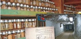
According to the conventional standards, keeping one mouse strain “in a living state” requires 10—12 pairs of stock animals and at least 50 young replacement animals. This number of animals requires approximately 3 m3 of vivarium space. While we deal with dozens of genotypes, this approach is still adequate; however, when there are thousands and hundreds of thousands of strains, the maintenance of mouse resources as breeding stocks becomes impossible. The way out of the dead-end is organization of cryoarchives.
Collections of genetic animal strains can be currently preserved as frozen embryos, spermatozoids, ova, and even ovarian tissue. However, the leading archives of genotypes focus on embryos. The embryo is a diploid organism, whereas spermatozoids and ova are haploid. Consequently, a full-fledged archiving of a genetic strain requires preservation of either embryos or gametes of both sexes. The former is more justified from a practical standpoint, as the latter implies one additional stage—fusing of thawed spermatozoids and ova to form embryos.
The embryo is an individual. Although this individual consists of only several cells, it already has its own developmental program encoded in the genome. Therefore, to restore a strain, it is enough just to transplant the thawed embryos into recipient females, where they will continue their development. The majority of experts agree that 200—500 embryos are enough to reliably preserve one strain. However, the world experience demonstrates that sometimes a strain can be revived from only 20 embryos.
Half a thousand embryos are easily packed in a volume of about 0.3 pint, which is by four orders of magnitude smaller than the space necessary for housing a strain of live mice. In addition, the cost of feed, litter, and maintenance is considerably higher than the expenses for cryopreservation (liquid nitrogen and its regular addition to a Dewar flask).
How long can be embryos preserved in a frozen state? Accumulated experience reliably demonstrates that in the case of an appropriately performed preservation, it does not matter how long the embryos have been stored in liquid nitrogen, be it several seconds or decades. For example, D. Whittingham, a British scientist, and his colleagues (in 1972, this particular team discovered the method for freezing embryos) continue today to produce mice using the embryos that they “canned” over 30 years ago: the embryos stored in the cryobank did not at all lose their ability “to revive.”
The story of how the first in the world (and still the largest one) cryobank of mouse genotypes with the Jackson Laboratory (USA) was set up is quite didactic. Its construction was initiated by the fire of 1947, which destroyed a considerable part of the inbred animal strains traditionally bred there. Consequently, as soon as the technology for cryopreservation of mouse embryos was developed, it was immediately implemented at this world genetic center. When this Laboratory was affected by an even more devastating fire in 1989 (the fire raged for over 5 hours and killed over half a million mice), the cryobank became a real fallback, as some strains existed only in the collection of this Lab and could be restored only from their own cryobank.
The example of the Jackson Laboratory was contagious, and cryoarchives appeared in other genetic centers; currently, their number reaches 20. The Japanese centers even have reserve banks in case of damage of the main cryobank. For example, the RIKEN BioResource Center has the main cryobank in Tsukuba and the reserve bank 400 km to the southwest (located in a facility with a synchrophasotron, constructed on a robust footing).
Moreover, the cryobanks are not only the way to overcome disasters. During traditional breeding, mutations inevitably accumulate in a genetic strain. In addition, mice, as any other living organisms, are sensitive to infections. Therefore, a cryobank of embryos is necessary not only for restoring a strain killed by infection or incident, but also for preserving its purity.
Pathogen free
More possibilities for creating live genetic “models” goes hand in hand with the tightening of the regulations for handling and manipulations with experimental animals. The requirement which is the most expensive and difficult to satisfy is providing the specific pathogen free (SPF) conditions for keeping animals.
Why do we need such animals? Getting rid of any infections eliminates the almost uncontrollable factor causing variation of experimental data; consequently, the reliability of results considerably increases. Therefore, GMP (good manufacturing practice) and GLP (good laboratory practice) standards demand that only SPF animals be used in the preclinical trials of drugs and assessment of biosafety of new materials, including foodstuffs.
Standardization of both housing conditions and animals themselves allows much smaller experimental groups. It is not accidental that the SPF animals in this country were for the first time used when the effect of space flight conditions on living organisms was studied — the necessity to decrease the number of experimental animals stemmed from the limitations imposed by the size of spacecraft.
In addition, a decrease in the number of experimental objects without a loss in experimental quality and improvement of animal housing conditions complies with the ethical requirements.
Another reason stimulating considerable efforts towards elimination of pathogens is an elevated sensitivity to diseases, characteristic of some genetic animal strains. In particular, solution of many immunological problems requires that individual components of the immune system be switched off, which makes animals unprotected from pathogens inevitable under natural conditions. An example is the above mentioned project on the mice producing human antibodies. An intermediate stage in creating such animals was the mice with their own immunoglobulin genes knocked out, i.e., the mice that are able to die of any, even benign infection.
Cryoarchives of experimental animal models are not only a backup for disasters, but also help maintain the genetic strains pureTo provide the SPF standard, laboratory animals are kept in clean rooms with a positive pressure formed by sterile air. The feed, litter, and cages are sterilized before they are conveyed to the clean zone of vivarium. In Japan, the feeds are decontaminated by gamma-irradiation, and the other components are passed through autoclaves. As mice, despite their small size, are not a nano-object, the volume of a cage for five—six animals is about 5 liters. Consequently, regular autoclaving of 1000—2 000 cages requires appropriate equipment capacities.
As a result, operation of animal breeding facilities in developed countries is provided by the industry that makes racks, cages, waterers, feeds, litter, and even special “toys” for mice. Comical as it may seem, these toys are a must in the United Kingdom and Scandinavian countries.
International cooperation
Rapidly increasing diversity of mouse genotypes has led to the foundation of an international organization for coordinating the activities of national centers for genetic resources. The Federation of International Mouse Resources (FIMRe) unites 16 Centers of North America, Europe, Asia, and Australia. The centers form collections of genotypes, making an agreement not to duplicate one another. These collections are supplemented by the genetic breeding and genetic engineer activities of the Centers themselves; however, the main contributors are departmental research laboratories.
The relations between the depositors to FIMRe cryoarchives and the customers are regulated by the material transfer agreements. The depositors acquire the following advantages:
• Guarantees of the author’s rights;
• Exemption from charges for storage and distribution of genetic model;
• Safe storage; and
• Last but not least—the citation index of depositors increases by over 88% (according to the data of the RIKEN BRC).
When the created genetic models are deposited, their departmental affiliation is taken into account. For example, the university and academic laboratories in Japan pay only the cost of animal breeding and housing; and commercial institutions pay 30% more. However, the animal sales proceeds in both cases do not cover the expenses for the genetic centers maintenance. Thus, the majority of genetic centers are funded from the state federal budgets of the countries that are really concerned about the development of high technologies for public health care.
The established network of FIMRe international centers has considerably contributed to the research into genetic bases of the organism functions in the norm and pathologies and the search for optimal prevention tools and drugs. Today, the following pattern is available for researchers to interact with the world genetic archives: formulating the need in experimental animals with given characteristics, searching for the required genotype in the FIMRe database, ordering the animals via the nearest FIMRe Center, obtaining the ordered strain by this Center, breeding these animals, and transferring them to the customer.
The current trend is a decrease in the exchange of pedigree animals versus an increase in the exchange of cryoproducts. It can be predicted that implementation of the KOMP-like projects will involve into the world turnover of model organisms the embryonic stem cell lines with specified genotype changes and then genetically engineered constructs: soon it will be possible to order them on application based on catalogs, as you can now order a chemical. This is especially efficient from the standpoint of biosafety, as even the most comprehensive control over the shipment of living organisms cannot rule out the risk of transborder exchange with undesirable microorganisms.
Alas, all these benefits of progress will be available only in the countries having their own Centers which can provide a complete cycle of manipulation with genetic resources, i.e., which have a cryobank, laboratories of reproductive technologies and transgenesis, and the SPF facilities for animal breeding and housing.
And in Russia?
In Russia, the only SPF animal breeding facility certified according to the international standards is in the Pushchino Scientific Center, located near Moscow. This Animal Breeding Facility with the Branch of Shemyakin and Ovchinnikov Institute of Bioorganic Chemistry, Russian Academy of Sciences, maintains about a dozen most “popular” mouse strains, several rat strains, and one strain of Syrian hamsters. Along with the reproduction of experimental objects, the Animal Breeding Facility is involved in cleanup of animals from pathogens, cryopreservation of sexual products and embryos, and surgical preparation of animals for chronic and acute experiments.
One of the main customers for SPF animals, the Laboratory of Biological Trials of the Pushchino Branch, is located in the immediate neighborhood. The availability of SPF animals allows this Institute to perform preclinical trials of drugs according to the GMP standard, ISO 9000, and, correspondingly, to get orders for such trials from foreign universities and pharmaceutical companies.
What is the situation to the east of the Ural Mountains? After Pushchino-on-Oka, the next center for breeding SPF animals is based in Japan. The vast territory between these geographical points is absolutely free of the specific pathogen free animals. It is a dismal fact as the Siberian Branch as well as the Ural and Far East Branches of the Russian Academy of Sciences (and the Russian Academy of Medical Sciences) are actively involved in research requiring laboratory mice and rats. Taking into account the corresponding departmental institutions as well as classical and medical universities, the number of potential corporate customers exceeds one hundred.
Currently, all these institutions are getting by on the limited set of genotypes available from the existing animal breeding facilities. However, this already interferes considerably with the basic research and classifies the results of applied research as “for house use only,” because the quality of experimental animals and their housing conditions do not meet the international standards.
As we consider our gloomy situation and bright prospects, we should note an important historical role of Academician V. K. Shumny, who five years ago, being Director of the Institute of Cytology and Genetics and Chairman of the United Scientific Council for Biological Sciences, managed to persuade the Russian Government to fund the construction of a state-of-the-art vivarium with elevated standards of animal housing.
It is not certain what particular reasons underlay the decision on constructing the SPF vivarium; however, this was undoubtedly hitting the bull’s eye. Using the artillery terms, we can state that this “shellfire” covered not only the targets visible at the moment when this decision was made, but also those that have become topical only now. During his recent visit to Germany, President of the Russian Federation D. Medvedev said again that “…Russia’s entry to the World Trade Organization is the gap that Russia should bridge”. In the context of our topic, this means that the international standards for all type of manipulations with laboratory animals should be introduced, which implies setting up an appropriate experimental base.
However, the strongest incentive to organization of the state-of-the-art center for live genetic models is the scientists’ desire to improve the reliability of their own research results. Indeed, an opportunity to work with comprehensively standardized experimental objects kept in a stringently controlled ambience in combination with an unlimited access to the world “genetic resources” is half the battle in the development of modern biological research, is not it?
This is even truer of the interdisciplinary studies aimed at solving basic problems related to the functions of living organisms at various organization levels. The corresponding solutions can answer the topical questions on the causes of diseases and aging, ways to success in various fields of competition, biological bases of social acceptance and rejection, and many others, making place for both a physicist and a lyrist. Development of the modern infrastructure will also be of great importance for training young scientists, as the opportunity to work at the world level (which implies the availability of experimental objects and necessary tools) is their most important motivation when they think what they should do with their lives.
This project will take to a new level research involving laboratory animals both within the Siberian Branch and Siberia on the whole (in particular, the international GMP and GLP standards will be met)Finally, one more motive to create a state-of-the-art genetic center was the regional program for development of the pharmaceutical industry in the Novosibirsk oblast (region), in particular, foundation of a pharmaceutical cluster comprising the Siberian Center for Pharmacology and Biotechnology (SCFB), institutes of the Siberian Branches of the Russian Academy of Sciences and Academy of Medical Sciences, and venture companies. This is a significant component of the overall innovation complex of the Novosibirsk Scientific Center and Novosibirsk oblast. Note that this research field related to development of new tools for disease diagnostics, prevention, and treatment is a hub where the interests of not only physicians, biologists, and chemists overlap, but also these of physicists involved in anything from nuclear physics to aerodynamics.
Undoubtedly, the issue of biomedical integration of natural sciences is a topic of a separate discussion. Note here that any works in this direction cannot do without the stage of preclinical trials, where the objects are experimental animals with specified genetic characteristics, whose quality and housing conditions should meet the stringent international standards.
Multifunctional and collective
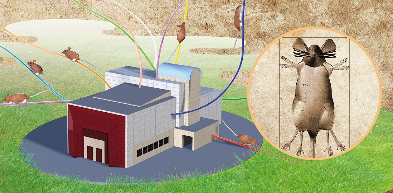
Thus, where are we today? According to the decision of the Russian Government, a state-of-the-art vivarium for small animals with elevated standards of animal housing is under construction with the Institute of Cytology and Genetics, Novosibirsk Scientific Center. Owing to its original architecture (the project leader is B. V. Nesterov, Leading Design and Research Institute of the Siberian Branch of the Russian Academy of Sciences), the building under construction is attracting everybody’s attention.
The total area of the vivarium is about 5,000 m2; 1,000 m2 of which meets the “clean room” standard (class C). The contractor is the Russian leader in his field, the Miass Plant of Medical Equipment. Two autonomous blocks for housing SPF laboratory animals are being assembled on the second and third floors. Four laboratory rooms with a total area 140 m2 are located on the first floor; the remaining space is allotted for various engineering and auxiliary facilities.
This design provides good opportunities for multifunctional use of this infrastructure object, and this is the only reasonable solution for the current situation. For example, the organization of the studies with laboratory animals in developed countries displays a historically established task sharing. An illustrative example is Japan, with its population close to that of Russia and its highest reputation in both hi-tech field in general and “health-efficient” technologies in particular. Many Japanese universities and commercial laboratories perform research with laboratory animals. All large universities have their own vivaria meeting the SPF standards and the laboratories producing transgenic mice. The university vivaria acquire most popular mouse and rat strains from specialized breeding facilities in addition to their own breeding activities. Specialized resource centers were organized in Japan for integration and preservation of a rapidly growing diversity of laboratory animals. An example is the RIKEN BioResource Center in Tsukuba, intended for mice and warehousing in its rapidly expanding cryoarchive over 3, 000 genetic strains. The National BioResource Project for the Rat with the Kyoto University as its leading institution is in charge of the rat strains (their number now is 458).
Note that an efficient operation of such infrastructure complexes providing both basic and applied research with laboratory animals requires all the above listed structural components as well as a number of auxiliary commercial organizations supplying the feed, litter, disinfectants, and other vitally necessary “little things”. As for us, we are completing the construction of the vivarium actually in the middle of an infrastructural semidesert. It is a semidesert as we have
• Vivaria located nearby, which, however, do not meet SPF housing conditions;
• Animal feeds, which, however, are not sterilized by gamma-irradiation; and
• A lot of trees (not surprising for Siberia!); though there is no sawdust of the quality necessary for litter.
Unfortunately, the list of the items we do not have at all could be much longer.
And yet, despite many, as we hope, transient shortcomings, we have no grounds for pessimism. The concept of this multifunctional complex was well received by the administration of the Institute of Cytology and Genetics, Academicians V. K. Shumny and N. A. Kolchanov, and then was developed collectively and approved by the scientific community of the Siberian Branch. This is demonstrated by the resolution of the General Meeting of the Siberian Scientific Center (no. 1 of November 15, 2007 and the Presidium of the Siberian Branch (no. 26 of January 24, 2008) on organization of the Shared Access Center with the SPF vivarium to provide for the research in the fields of genetics, molecular biology, physiology, biomedicine, nanobiosafety, and pharmacology. The project was also presented and approved at the session of the Presidium of the Siberian Branch of the Russian Academy of Medical Sciences (February 20, 2008).
Quantum Sufficit!
According to the concept of a multifunctional complex, the new Center will include:
• Cryobank and laboratory of reproductive technologies intended for creating the archive of mouse and rat genetic strains via cryopreservation of sexual cells, early embryos, and embryonic stem cells; for adjusting the unique animals strains bred in Russia to the SPF standard with an aim to organize an exchange with cryoarchives in Russia and abroad, first and foremost, with FIMRe genetic centers; for elaboration of new cryopreservation technologies for various animals species; and for creation of cryoarchives for unique breeds of agricultural animals;
• Breeding sector for breeding and maintaining the pedigree stocks of various mouse and rat strains;
• Sector of transgenic animals for genetic engineering creation of new animal strains with specified genetic characteristics;
• Laboratories of physiological and ethological phenotyping for intravital examination of animal behavior and morphophysiological processes within the SPF zone;
• Sample processing laboratories for the initial processing of samples taken from experimental animals; and
• Information Support Center, which would provide an automated control of all types of manipulations with animals and connection with the databases of world centers for laboratory animal bioresources.
When completed, this project will dramatically advance (in particular, the international GMP and GLP standards will be met) research connected with laboratory animals in the Siberian Branch and all over Siberia. We will be able, at last, to reject the “orphan ideology”, which in this sphere of science organization shows in the standard question on how many mouse strains will be kept in the new vivarium. The only correct answer today is “As many as you need and of the genotypes necessary for you to solve your specific problems”.
This Center will considerably improve the facilities for intravital morphofunctional examination, as the technical maintenance in this field has considerably advanced due to fMRI tomography, IR imaging, telemetry, intravital IR scanning, and so on. These approaches are most important when studying the changes both occurring during ontogenesis and induced by various experimental impacts, for example, in preclinical trials of new tools for disease prevention and treatment, biosafety assessment of new nanomaterials, etc.
Certainly, skeptics can doubt the feasibility of these plans. However, the experience of successfully developing countries, such as South Korea, China, and India, suggests that one should either burst into the sphere of new technologies, or leave them alone and continue to drill wells…
The fact that the material resources allocated for the construction of the new Center go hand in hand with proper intellectual and professional support of the future activities also adds optimism. Though we have no cryobank yet, we have S. Ya. Amstislavsky, who, in the absence of his own laboratory facilities, is working in Finland on expanding the list of species suitable for cryopreservation (including European mink, whose preservation is within the national liability of the Russian Federation). In addition, the Institute of Cytology and Genetics has accumulated considerable experience in breeding experimental animals with genetic predisposition to several diseases.
The genetic engineering research performed (frequently, in collaboration) at this Institute and at the Institute of Chemical Biology and Fundamental Medicine, Siberian Branch of the Russian Academy of Sciences, suggests that the future genetic collection of the new Center will be successfully replenished with its own transgenic animals created to match the specific problems the Siberian science will be dealing with. This will be done based on the interinstitute collaboration in transgenesis, with the SPF vivarium involved in some of these works.
The opportunity to perform research with comprehensively standardized experimental objects kept in a stringently controlled ambience in combination with an unlimited access to the world “genetic resources” is half the battle in the development of modern biological researchToday, Siberian Branch uses state-of-the-art methods to study molecular genetic, physiological, morphological, and behavioral processes in living organisms. For example, the approaches to intravital monitoring of such processes developed at the Tomography Center are used in joint research with the Institute of Cytology and Genetics and other biological institutes. One of the most successful laboratories (Institute of Chemical Biology and Fundamental Medicine) involved in pharmacological research provides the connection between chemical science and practical medicine (for details, see further issues of the journal).
The large-scale and manifold research into living organisms with the help of radiation technologies is conducted in collaboration with the Institute of Nuclear Physics and Institute of Chemical Kinetics and Combustion. We hope that along with basic problems, these experts will assist in solving the practical problem of irradiation sterilizing, necessary for raising SPF animals.
From structure to function
James Watson, a Nobel laureate, wrote in his memoirs that had commenced his scientific career as an ornithologist and ever since had been most curious to know how the intricate ornaments decorating males of many avian species were stably repeated in the line of generations. Together with Francis Crick, he considerably contributed to the solution of this problem by deciphering the DNA structure. Now that genome sequences of humans and many other species have been completely determined, we have all theoretical and technological prerequisites for the next step in our aspiration to understand how genetic information is transformed into the “life histories” of individuals.
This basic problem is not easy to solve. It is more probable that the solution will form gradually as an integrated result of a tremendous number of specific pieces of knowledge about the “trajectories” leading from a gene to the trait or from a genotype to the phenotype. The obligatory element in this “technological” cycle of understanding the living systems (from morphological description to physiological explanation) is experiments on animals with given genetic characteristics. Indeed, the constructions consolidating the results of analysis of nucleotide sequences, studies of the structures and functions of biomolecules, detailing of cell structures, mathematical modeling, etc., can be ultimately verified only at the level of the entire organism. Recollect the famous movie “The Meeting Place Cannot Be Changed”, a Soviet cult movie: a piano player can be recognized from the shape of his hands or an ear to music, but it is better just to ask him to play…
The need to accumulate specific pieces of knowledge is dictated not only by an unimaginable complexity of living organisms, but also by the lack of “unified structures” for each biological species, including humans. Therefore, biomedical research aimed at searching for the methods of treating “patients rather than diseases” involves an ever increasing number of various living genetic “models”.
One of the stimuli inducing elaboration of such models is an avalanche of information about the genes associated with particular pathologies. However, only in very rare cases, researchers succeed in performing a complete cycle of studies comprising identification of the genotypes connected with a disease, creation of the corresponding model animals, analysis of the mechanisms of genetically determined pathogenesis, and search for adequate correction tools.
Note that the frequencies of the genes associated with a disease and pattern of these associations have specific ethnic features. Thus, provision of conditions for implementing this set of studies is among the national priorities, as the saying goes, “It’s up to you to have a boat, if you want to stay afloat”. This is reflected in a rapidly increasing number of such genetic centers in all developed countries. Expansion of the range of model mammals with specified genetic characteristics is in the tideway of general trends in the development of modern biology and medicine.
Note that establishment of the Center that will provide access to the world archives of laboratory animals also has far-reaching consequences, as the development of the infrastructure itself has a tremendous influence on the raising of a competitive young generation. This applies to any forms of competitive activities; for example, the love for tennis of the first Russian President has resulted in improved conditions for young players’ training, and our tennis players now head the world list.
In science, it is even more important than in sports that the talented youth work in their motherland. And they should choose to stay not because of visa denial but because the conditions of their life and work here will be at the European level. The new Center located in Akademgorodok will undoubtedly help grow highly professional experts in various fields of biology and biotechnology from students of Novosibirsk State University and young researchers of academic institutes with the Siberian Branches of the Russian Academy of Sciences and Academy of Medical Sciences.
Thus, the SPF vivarium with the Institute of Cytology and Genetics, which is under construction thanks to the start capital granted by the Government of the Russian Federation, is bound to become a center that integrates the research activities of scientists who need experimental animal models. It must become a shared access center, as it is necessary for many institutes of the Siberian Branches in various regions of the Urals, Siberia, and Far East; and for classical, medical, and agricultural universities. This interest shows already as tangible scientific, methodical, and financial support under the program ”Genomics, Proteomics, and Bioinformatics”, headed by Academicians R. Z. Sagdeev, V. V. Vlassov, and N. A. Kolchanov.
Development of genetic resources centers, which in industrially developed countries is on the list of national priorities, will contribute to solving not only basic problems, but also to one of the main problems faced by a state—preservation of national healthIn the developed countries, such centers are on the list of national priorities and can be regarded as a symbol of statehood along with the hymn, emblem, and flag. There is nothing surprising about it: such genetic centers are an indispensable element of the science and technology complex designed to solve one of the main problems a country faces—to contribute to the health of the nation through improving the health of its citizens.
References:
Abbott А., Harbor В. Genetists prepare for deluge of mutant mice // Nature.—2004.—N. 432.—541 р.
Collins F. S. et al. The International knockout mouse consortium // Cell. 2007.—N. 128.—Рp. 9—13.
Davisson M. FIMRe: Federation of International Mouse Resources: global networking of resource centers // Mamm. Genome.—2006.—N. 17 (5).—363 р.
Gachon F. et al. The loss of circadian PAR bZip transcription factors results in epilepsy // Genes. & Dev.—2004.—N. 18.— Рp. 1397—1412.
Sprengel R., Hasa M. T. Tetracycline-controlled genetic switches // HEP.—2007.—№ 178.—Рp. 49—72.
Xiao-Wei Tan et al. Fetal microchemerism in the maternal mouse brain: a novel population of fetal progenitor or stem cells able to cross blood-brain barrier? // Stem. Cells.—2005.— N. 23. — Рp. 1443—1452.
* This and the following sections were written together with S.Ya. Amstislavsky


